Creative work the story of the discovery of the cell
MUNICIPAL BUDGETARY EDUCATIONAL INSTITUTION "PODBELEVSKAYA SECONDARY SCHOOL"
THE HISTORY OF THE DISCOVERY OF THE CELL
Completed by: Aleshkina Nadezhda Vladimirovna,
5th grade student
Head: Krasnoshchekova Irina Nikolaevna,
chemistry and biology teacher
2016
Table of contents ( content) p.
Introduction …………………………………………………………………… .3
Chapter 1. History of the invention of the microscope …… ... ……………………… 3
Chapter 2. History of cell opening …… .. ……………………………… ..5
Practical part… .. ………………………………………………… .9
Conclusions …… ... ……………………………………………………………… ..10
Used literature ……. …………………………………………… 11
INTRODUCTION
I'm in the 5th grade. This year we started to study a new subject - biology. Biology is the science of living nature. Biologists study the diversity of living things, the structure of their bodies and the work of various organs, the reproduction and development of organisms, their relationship with each other and with inanimate nature.
All living things have a cellular structure. Among them are unicellular and multicellular organisms. Most living cells have three main parts: the membrane, the cytoplasm, and the nucleus.
In one of the biology lessons, we examined ready-made micropreparations of various cells under a microscope. In extracurricular activities (Secrets of the microworld), we examined the infusoria-shoe, watched how it moves, we ourselves prepared micropreparations from onion skin and tomato pulp. And in them we were able to see the nucleus, cytoplasm and envelope.
I wondered how humanity managed to find out what organisms are made of, and how it became possible to see a cell.
Purpose of work:find out how the invention of the microscope influenced the discovery of the cell.
Tasks:
- to study the history of the invention of the microscope;
- study the history of the opening of the cell;
- conduct a survey;
- to make an experiment;
- draw conclusions .
Object of study : cell
Subject of study : opening the cell
Methods used : analysis, experiment, observation, conclusions.
Chapter 1. HISTORY OF THE INVENTION OF THE MICROSCOPE
The invention of the microscope, a device so important for the whole of science, is primarily due to the influence of the development of optics. Some of the optical properties of curved surfaces were already known to Euclid (300 BC) and Ptolemy (127-151), but their magnifying ability has not found practical application. In this regard, the first glasses were invented by Salvinio delhi Arleati in Italy only in 1285. In the 16th century, Leonardo da Vinci and Maurolico showed that it is better to study small objects with a magnifying glass.
Rice. 1. The first microscope
The first microscope was created only in 1595 by Zacharius Jansen (Jansen). The invention consisted in the fact that Zacharius Jansen mounted two convex lenses inside one tube, thereby laying the foundation for creating complex microscopes. Focusing on the object under study was achieved by means of a retractable tube. The microscope magnification ranged from 3 to 10 times. And this was a real breakthrough in the field of microscopy! Each of his next microscope, he significantly improved.
During this period (16th century), Danish, English and Italian research instruments gradually began their development, laying the foundation for modern microscopy.
The rapid spread and improvement of microscopes began after Galileo, improving the telescope designed by him, began to use it as a kind of microscope (1609-1610), changing the distance between the objective and the eyepiece.
Later, in 1624, having achieved the manufacture of short-focus lenses, Galileo significantly reduced the size of his microscope.
In 1625, a member of the Roman "Academy of the Vigilant" I. Faber proposed the term"microscope"... The first advances in the use of the microscope in scientific biological research were achieved by Hooke, who was the first to describe the plant cell (around 1665). In his book, Micrographia, Hooke described the construction of a microscope. From now on a new world of living beings was revealed, more diverse and infinitely more original than the world we see.(http://www.vita-club.ru/ micros1.htm)
Chapter 2. HISTORY OF DISCOVERY OF THE CELL
A cell is an elementary structural and functional unit of an organism, possessing all the basic characteristics of a living thing. Cells are able to multiply, grow, exchange matter and energy with the environment, and respond to changes in this environment. Each cell contains hereditary material, which contains information about all the signs and properties of a given organism.( )
R 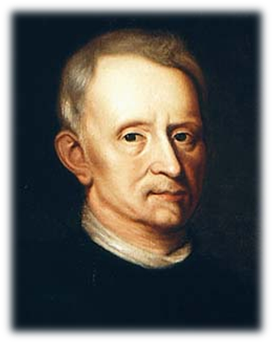 is. 2. Robert Hooke.
English scientist Robert Hooke (1635-1703) saw plant cells for the first time.
It happened in 1665. It was like this: Hooke examined a thin section of a linden cork with a magnification of 30 times. He discovered that the cork was made up of many small cavities, chambers, which he called "cells." It was he who introduced the concept of "cell" into science.(Pleshakov A.A., Vvedensky E.L. Biology. Introduction to biology: a textbook for grade 5 of general education institutions / M.: OOO "Russian word - textbook", 2014.)True, these were not living cells, but already dead cells. Hooke believed that the cells themselves are emptiness, and the contents of a living organism are enclosed in a frame (cell wall).
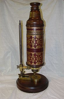
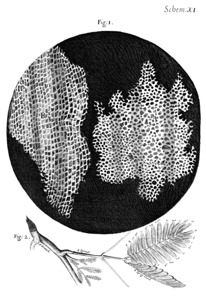
Fig. 3 R. Hooke's microscope Fig. 4... Cork cells studied by Robert Hooke
Soon the cellular structure of plants was confirmed by the Italian physician and microscopist M. Malpighi and the English botanist N. Gru. Their attention was attracted by the shape of the cells and the structure of their membranes. As a result, the concept of cells was given as "bags" or "bubbles" filled with "nutritional juice". ( )
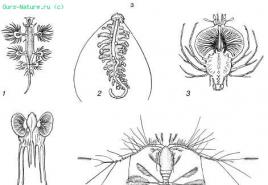
DutchmanAntonia WangLevengukdescribed the amazing miracles that he discovered with his microscope in a drop of water, in an infusion of pepper, in the mud of a river, in the hollow of his own tooth. Levenguk using a microscope discovered and sketched spermatozoa, various protozoa, details of the structure of bone tissue (1673-1677).
“With the greatest amazement I saw in the drop a great multitude of animals moving briskly in all directions, like a pike in water. The smallest of these tiny animals is a thousand times smaller than the eyes of an adult louse. "
Levenguk's best loops were magnified 270 times. With them, he saw for the first time blood corpuscles, the movement of blood in the capillary vessels of the tail of a tadpole, and striped muscles. He opened the ciliates. He first plunged into the world of microscopic unicellular algae, where the border between animal and plant lies; where a moving animal, like a green plant, possesses chlorophyll and feeds by absorbing light; where the plant, still attached to the substrate, has lost chlorophyll and swallows bacteria. Finally, he even saw bacteria in great variety. But, of course, then there was still no remote possibility to understand either the significance of bacteria for humans, or the meaning of the green substance - chlorophyll, or the border between plant and animal. ( )
The description of these "animals" ("animals"), as he called them, earned the Dutchman world fame. But most importantly, Levenguk's discoveries aroused interest in the study of the living microworld.(Encyclopedia for children. Vol.2 Biology. -5th ed. / Chief editor M.D. Aksenova. - M .: Avanta +, 2001)
R 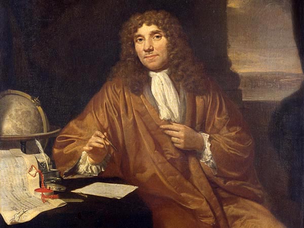 fig.5 by Antonia Van Leeuwenhoek
In 1693, during the stay of Peter I in Delphi, A. Levenguk demonstrated to him how blood moves in the fin of a fish. These demonstrations made such a great impression on Peter I that, upon returning to Russia, he created a workshop for optical instruments. In 1725, the St. Petersburg Academy of Sciences was organized. The talented masters I.E. Belyaev, I.P. Kulibin made microscopes, in the design of which academicians L. Euler, F. Epinus took part.
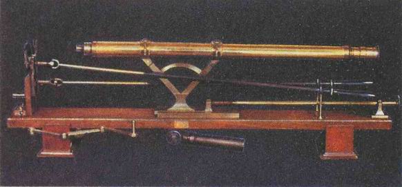
Fig. 6 Microscope made by Russian craftsmen.
For a long time, however, the microscope remained more of an expensive toy than a scientific instrument. Only in the 30s.XIXv. the lenses have been improved to such an extent that they were able to provide strong magnification and image clarity. Biologists were able to consider that each cell is covered with a membrane, and under it there is a liquid with a nucleus. The nucleus in plant cells was first described in 1831 by Scottish botanist Robert Brown.
The famous German biologist Theodor Schwann (1810-1882) was the first scientist to understand that the cell is the smallest element that makes up all tissues of animal organs. Later, on the basis of his own research and the works of the German botanist Matthias Jakob Schleiden (1804-1881), Schwann came to the conclusion that the law of cell structure is also valid for plants. In 1839 he published his later famous essay "Microscopic Investigations of Correspondence in the Structure and Growth of Animals and Plants."
(Encyclopedia for children. Vol.2 Biology. -5th ed. / Chief editor M.D. Aksenova. - M .: Avanta +, 2001)
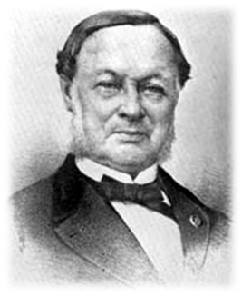
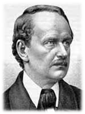
Fig. 7 Theodor Schwann Fig. 8 Matthias Jakob Schleiden
T. Schwann and M. Schleiden made a number of generalizations, which were later calledcell theory :
All living things are made of cells;
The cells of plants and animals have a similar structure;
Each cell is capable of independent existence;
The body's activity is the sum of the vital processes of its constituent cells.
They mistakenly believed that cells in the body arise from non-cellular matter. An important addition to the cell theory was Rudolf Virchow's principle: "Every cell is from a cell" (1859). ( )
PRACTICAL PART
The first part of my research was the questionnaire. I interviewed 60 people, these are students of our school and residents of the village of Podbelevets. The first question on my questionnaire was: Do you know that all organisms are made up of cells? 59 people (98.3%) know the answer to this question. Almost all survey participants (58 people - 96.6%) know that a cell can be seen under a microscope. The majority (53 people - 88.3%) called the nucleus the main part of the cell and answered correctly, 2 people (3.3%) - the cytoplasm, 2 people (3.3%) - the membrane, and 3 people (5%) do not know the answer to this question. Robert Hooke was called the discoverer of the cell by 23 people (38.3%), and this is the correct answer. 19 people (31.6%) named Levenguk, 3 people (5%) - Schwann and Schleiden, and 15 people (25%) found it difficult to answer.
According to the results of the survey, we can say that the majority of the respondents have an idea about the cell and the methods of studying it. Not everyone knows the story of the cell's discovery. Primary school students do not know the answers to many questions, but they still have more to come.
The second stage of my work was an experiment. I prepared a micropreparation of a plant cell of an onion skin and examined it under a microscope. I saw many cells in which I identified three main parts of the cell: the nucleus, the cytoplasm and the membrane.
Method n preparation of onion skin micropreparation.
Separated a fleshy scale from a piece of onion. On the inside there is a thin film. She took it off with tweezers.
I put it on a glass slide, dropped a drop of iodine solution and covered it with a cover glass.
Examined the micropreparation under a microscope under low and high magnification.
The modern school microscope is simple, and allows students to independently work with it, to conduct small studies. It is not difficult for me to prepare micropreparations of plant and animal cells myself and examine them under a microscope.
CONCLUSION
Having studied the literature on this issue, I found out that the microscope was invented at the end of the 16th century (1595) by a Dutch eyeglass masterZacharius Jansen(Jansen). Using a microscope, the English scientist Robert Hooke (1665) discovered cells when examining a linden cork and called them cells. Dutchman Antonia Van Leeuwenhoek improved the microscope and described blood cells, spermatozoa, some unicellular animals, etc. Scottish botanist Robert Brown (1831) discovered a dense formation inside the cell, which he called the nucleus. In 1838, German scientists Theodor Schwann and Mathias Schleiden created the cell theory. They noted that all plant and animal organisms are composed of cells that are similar in structure. In 1858, the German scientist Rudolf Virchow made an addition to the cell theory, indicating that the cell comes from the cell.
Thus, based on my research, the following conclusions can be drawn.
The invention and improvement of the microscope allowed humanity to look into the microscopic world of living.
With the help of a microscope, it became possible not only to see the cell and its main parts, but also to study its vital activity.
Based on the results of the survey, I found out that the majority of the surveyed population, regardless of age, are interested in biology, they know its basics.
USED BOOKS
Belyaev D.K., Borodin P.M., Vorontsov N.N. et al. General biology: textbook. for 10-11 grades of general educational institutions / - M .: Education 2005.
A.A. Kamensky General biology. Grade 10-11: textbook for general education institutions / - M .: Bustard 2012
Pleshakov A.A., Vvedensky E.L. Biology. Introduction to biology: a textbook for grade 5 general education. institutions: line "Rakurs" / A.A. Pleshakov, E.L. Vvedensky, - M .: LLC "Russian word - textbook", 2013.
Teremov A.V. Biology. General patterns of life: 9kl .: textbook for students of general education institutions / A.V. Teremov, R.A. Petrosova, A.I. Nikishov.- M .: Humanities. publishing center VLADOS, 2015.
Encyclopedia for children. Vol.2 Biology. -5th ed. / Chief ed. M.D. Aksenova. - M .: Avanta +, 2001.
Internet sites