Epithelial and connective tissues
1. Fabric classification
Textile - This is a historically (phylogenetically) established system of cells and non-cellular structures with a common structure, specializing in the performance of certain functions.
Each tissue originates from a specific germ layer and consists of cells and non-cellular matter.
In the animal body, several types of tissues are distinguished: epithelial, connective, support-trophic, muscle and nervous tissues.
2. Types of epithelial tissues. Their structure and functions
Epithelial tissues or epithelium line the surface of the body, serous membranes, the inner surface of hollow organs (stomach, intestines, bladder) and form most of the body's glands. They originated from all three germ layers - ectoderm, endoderm, mesoderm.
Epithelium is a layer of cells located on the basement membrane, under which lies loose connective tissue. There is almost no intermediate substance in the epithelium and the cells are in close contact with each other. Epithelial tissues do not have blood vessels and their nutrition is carried out through the basement membrane from the side of the underlying connective tissue. Fabrics have a high regenerative capacity.
The epithelium has a number of functions:
- Protective - protects other tissues from environmental influences. This function is characteristic of the epithelium of the skin;
- Nutrient (trophic) - absorption of nutrients. This function is performed, for example, by the epithelium of the gastrointestinal tract;
- Excretory - removal of unnecessary substances from the body (CO 2, urea);
- Secretory - most of the glands are built from epithelial cells.
Epithelial tissues can be classified in the form of a diagram. Monolayer and stratified epithelium differ in cell shape (Fig. 1).
Single layered, squamous epitheliumconsists of flat cells located on the basement membrane. This epithelium is called mesothelium and lines the surface of the pleura, pericardial sac and peritoneum.
Endothelium is a derivative of the mesenchyme and is a continuous layer of flat cells covering the inner surface of the blood and lymphatic vessels.
Single layered cuboidal epitheliumlines the tubules of the kidney, which excrete the ducts of the glands.
Single layered columnar epitheliumcomposed of prismatic cells. This epithelium lines the inner surface of the stomach, intestines, uterus, oviducts, renal tubules. Goblet cells are found in the intestinal epithelium. These are unicellular glands that secrete mucus.
In the small intestine, epithelial cells have a special formation on the surface - a border. It consists of a large number of microvilli, which increases the surface of the cell and promotes better absorption of nutrients and other substances. The epithelial cells lining the uterus have ciliated cilia and are called ciliated epithelium.
Single layered epitheliumdiffers in that its cells have a different shape and, as a result, their nuclei lie at different levels. This epithelium has ciliated cilia and is also called ciliated. It lines the airways and some parts of the reproductive system. The movement of the cilia removes dust particles from the upper respiratory tract.
Stratified squamous epitheliumis a relatively thick layer consisting of many layers of cells. Only the deepest layer is in contact with the basement membrane. Stratified epithelium performs a protective function and is divided into keratinized and non-keratinized.
non-keratinizingThe epithelium lines the surface of the cornea of the eye, oral cavity and esophagus. Consists of cells of various shapes. The basal layer consists of cylindrical cells; then cells of various shapes with short thick processes are located - a layer of spiny cells. The uppermost layer consists of flat cells, gradually dying and falling off.
keratinizing The epithelium covers the surface of the skin and is called the epidermis. It consists of 4-5 layers of cells of different shapes and functions. The inner layer, basal, consists of cylindrical cells capable of reproduction. The layer of spiny cells consists of cells with cytoplasmic islands, with the help of which the cells come into contact with each other. The granular layer consists of flattened cells containing granules. The shiny layer in the form of a shiny ribbon, consists of cells, the boundaries of which are not visible due to the shiny substance - eleidin. The stratum corneum consists of flat scales filled with keratin. The most superficial scales of the stratum corneum gradually fall off, but are replenished by multiplying cells of the basal layer. The stratum corneum is characterized by resistance to external, chemical influences, elasticity and low thermal conductivity, which ensures the protective function of the epidermis.
transitional epitheliumcharacterized by the fact that its appearance varies depending on the state of the organ. It consists of two layers - basal - in the form of small flattened cells and integumentary - large, slightly flattened cells. The epithelium lines the bladder, ureters, pelvis, renal calyces. When the organ wall contracts, the transitional epithelium looks like a thick layer in which the basal layer becomes multi-row. If the organ is stretched, the epithelium becomes thin and the shape of the cells changes.
3. Connective tissues (except: blood and lymph). Their structure and functions
Connective tissues are diverse in their structure, as they perform supporting, trophic and protective functions. They consist of cells and intercellular substance, which is more numerous than cells. These tissues have a high regenerative capacity, plasticity, adaptation to changing conditions of existence. Their growth and development occurs due to reproduction, transformation of poorly differentiated young cells.
Connective tissues originated from the mesenchyme, i.e. embryonic connective tissue, which was formed from the middle germ layer - the mesoderm.
There are several types of connective tissue:
- Blood and lymph;
- Loose fibrous unformed tissue;
- Dense fibrous (formed and unformed) tissue;
- reticular tissue;
- fatty;
- cartilaginous;
- Bone;
Of these types, dense fibrous, cartilage and bone perform a supporting function, the rest of the tissues are protective and trophic.
Loose fibrous irregular connective tissue
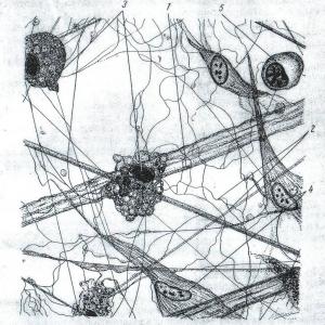
This tissue consists of various cellular elements and intercellular substance (Fig. 2). It is part of all organs, in many of them it forms the stroma of the organ. It accompanies the blood vessels, through it there is an exchange of substances between the blood and the cells of organs and, in particular, the transfer of nutrients from the blood to the tissues.
The intercellular substance includes three types of fibers: collagen, elastic and reticular. Collagen fibers are located in different directions in the form of straight or wave-like curved strands with a thickness of 1-3 microns or more. Elastic fibers are thinner than collagen fibers, they anastomose with each other and form a more or less broadly braided network. The reticular fibers are thin, forming a delicate mesh.
The ground substance is a gelatinous, structureless mass that fills the space between the cells and fibers of the connective tissue.
The cellular elements of loose fibrous tissue include the following cells: fibroblasts, macrophages, plasma, mast, fat, pigment and adventitial cells.
fibroblasts - These are the most numerous flat cells that have a spindle shape on the cut, often with processes. They are capable of reproduction. They take part in the formation of the ground substance, in particular, they form connective tissue fibers.
Macrophages - cells capable of absorbing and digesting microbial bodies. There are macrophages that are in a calm state - histocytes and wandering - free macrophages. They can be round, elongated and irregular in shape. They are capable of amoeboid movements, destroy microorganisms, neutralize toxins, participate in the formation of immunity.
Plasma cellsfound in the loose connective tissue of the intestine, lymph nodes, bone marrow. They are small, round or oval in shape. They play an important role in the body's defense reactions, for example, they take part in the synthesis of antibodies. They produce blood globulins.
mast cells - in their cytoplasm there is granularity (granules). They are found in all organs where there is a layer of loose, unformed connective tissue. The form is varied; granules contain heparin, histamine, hyaluronic acid. The value of cells lies in the secretion of these substances and the regulation of microcirculation.
fat cells - These are cells capable of depositing reserve fat in the form of droplets in the cytoplasm. They can crowd out other cells and form adipose tissue. Cells are spherical.
adventitial cellslocated along the course of blood capillaries. They have an elongated shape with a core in the center. Capable of reproduction and transformation into other cellular forms of connective tissue. When a number of connective tissue cells die, their replenishment occurs due to these cells.
Dense fibrous connective tissue
This fabric is divided into dense shaped and unshaped.
Thick loose fabricconsists of a relatively large number of densely packed connective tissue fibers and a small number of cellular elements between the fibers.
Thick woven fabriccharacterized by a certain arrangement of connective tissue fibers. Tendons, ligaments and some other formations are built from this tissue. Tendons are composed of tightly packed parallel bundles of collagen fibers. Between them there is a thin elastic network and small spaces are filled with the main substance. Of the cellular forms in the tendons, there are only fibrocytes.
A type of dense connective tissue iselastic fibrous connective tissue.Some cords are built from it, for example, vocal cords. In these ligaments, thick rounded or flattened elastic fibers are arranged parallel side by side, but often branch. The space between them is filled with loose unformed connective tissue. Elastic tissue forms a shell of round vessels, is part of the walls of the trachea and bronchi.
cartilage tissue
This tissue consists of cells, a large amount of intercellular substance and performs a mechanical function.
There are two types of cartilage cells:
- Chondrocytes are oval cells with a nucleus. They are located in special capsules surrounded by intercellular substance. Cells are located alone or in 2-4 cells or more, they are called isogenic groups.
- Chondroblasts - These are young, flattened cells located on the periphery of the cartilage.
There are three types of cartilage: gliane, elastic and collagen.
Glan cartilage. It occurs in many organs: in the ribs, on the articular surfaces of bones, along the airways. Its intercellular substance is homogeneous and translucent.
Elastic cartilage. In its intercellular substance there are well-developed elastic fibers. The epiglottis, cartilages of the larynx are built from this tissue, and it is part of the wall of the external auditory canals.
collagen cartilage.Its intermediate substance consists of dense fibrous connective tissue, i.e. includes parallel bundles of collagen fibers. Intervertebral discs are built from this tissue; it is found in the sternoclavicular and mandibular joints.
All types of cartilage are covered with dense fibrous tissue, in which collagen and elastic fibers are found, as well as cells similar to fibroblasts. This tissue is called the perichondrium; richly supplied with blood vessels and nerves. Cartilage grows at the expense of the perichondrium by transforming its cellular elements into cartilage cells. There are no vessels in the intercellular substance of mature cartilage and its nutrition occurs by diffusion of substances from the vessels of the perichondrium.
Bone
This tissue consists of cells and a dense intercellular substance. It differs in that its intercellular substance is calcified. This gives the bone the hardness needed to perform its supporting function. The bones of the skeleton are built from this tissue.
The cellular elements of bone tissue include bone cells, or osteocytes, osteoblasts and osteoclasts.
Osteocytes - have a process shape and a compact, dark-colored nucleus. The cells lie in bone cavities that follow the contours of osteocytes (Fig. 3). Osteocytes are incapable of reproduction.
osteoblasts - Cells that make bone. They are rounded, sometimes contain several nuclei, are located in the periosteum.
osteoclasts - cells that take an active part in the destruction of calcified cartilage and bone. These are multinucleated, rather large cells. Throughout life, the destruction of the structural parts of the bone tissue occurs and at the same time the formation of new ones, both at the site of destruction and from the side of the periosteum. Osteoclasts and osteoblasts take part in this process.
intercellular substancebone tissue consists of an amorphous ground substance in which ossein fibers are located. There are coarse fibrous tissue, which is present in embryos, and lamellar bone tissue, which is present in adults and children.
The structural unit of bone tissue isbone plate.It is formed by bone cells lying in capsules and fine-fibrous intercellular substance impregnated with calcium salts. Ossein fibers of these plates lie parallel to each other in a certain direction. In neighboring plates, the fibers usually have a direction perpendicular to them, which ensures greater strength of the bone tissue. Bone plates in different bones are arranged in a certain order. Almost all flat, tubular and mixed bones of the skeleton are built from them.
In the diaphysis of the tubular bone, the plates form complex systems in which three layers are distinguished: 1) the outer one, in which the plates do not form complete rings and overlap on the surface with the next layer of plates; 2) the middle layer is formed by osteons. In the osteon, the bone plates are arranged concentrically around the blood vessels (Fig. 4); 3) the inner layer of the plates delimits the bone marrow space, where the bone marrow is located.
The bone grows and regenerates due to the periosteum, which covers the outer surface of the bone and consists of fine fibrous connective tissue and osteoblasts.
4. Structure and functions of nervous and muscular tissues
These tissues are referred to as excitable tissues, i.e. they are capable of responding to irritation with excitation and conducting it at a distance.
Muscle tissues
By origin and structure, muscle tissues differ significantly from each other, but they are united by the ability to contract, which ensures the motor function of organs and the body as a whole. The muscle elements are elongated and connected either with other muscle elements or with supporting formations.
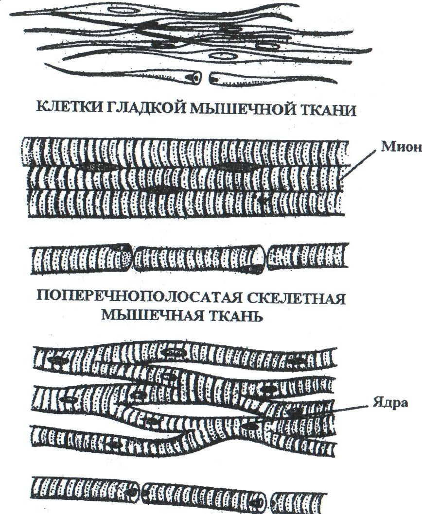
There are smooth, striated muscle tissue and muscle tissue of the heart (Fig. 5).
Smooth muscle tissue.
This tissue is formed from mesenchyme. The structural unit of this tissue is a smooth muscle cell. It has an elongated fusiform shape and is covered with a cell membrane. These cells are tightly adjacent to each other, forming layers and groups, separated from each other by a loose, unformed connective tissue.
The cell nucleus has an elongated shape and is located in the center. Myofibrils are located in the cytoplasm, they go along the periphery of the cell along its axis. They consist of thin threads and are the contractile element of the muscle.
Cells are located in the walls of blood vessels and most of the internal hollow organs (stomach, intestines, uterus, bladder). Smooth muscle activity is regulated by the autonomic nervous system. Muscle contractions do not obey the will of a person and therefore smooth muscle tissue is called involuntary muscles.
Striated muscle tissue.
This tissue was formed from myotomes, derivatives of the mesoderm. The structural unit of this tissue is the striated muscle fiber. This cylindrical body is a symplast. It is covered with a membrane - sarcolemma, and the cytoplasm is called - sarcoplasm, in which there are numerous nuclei and myofibrils. Myofibrils form a bundle of continuous fibers running from one end of the fiber to the other parallel to its axis. Each myofibril consists of discs that have a different chemical composition and appear dark and light under a microscope. Homogeneous discs of all myofibrils coincide, and therefore the muscle fiber appears to be striated. Myofibrils are the contractile apparatus of the muscle fiber.
All skeletal muscles are built from striated muscle tissue. Musculature is arbitrary, because. its contraction may occur under the influence of neurons in the motor cortex of the cerebral hemispheres.
Muscular tissue of the heart.
Myocardium - the middle layer of the heart - is built from striated muscle cells (cardiomyocytes). There are two types of cells: typical contractile cells and atypical cardiac myocytes, which make up the conduction system of the heart.
Typical muscle cells perform a contractile function; they are rectangular in shape, in the center there are 1-2 nuclei, myofibrils are located along the periphery. There are intercalated discs between adjacent myocytes. With their help, myocytes are collected into muscle fibers, separated from each other by fine-fibred connective tissue. Connecting fibers pass between adjacent muscle fibers, which provide contraction of the myocardium as a whole.
The conduction system of the heart is formed by muscle fibers, consisting of atypical muscle cells. They are larger than contractile ones, richer in sarcoplasm, but poorer in myofibrils, which often intersect. The nuclei are larger and not always in the center. The fibers of the conducting system are surrounded by a dense plexus of nerve fibers.
nervous tissue.
Nervous tissue consists of nerve cells with a specific function, and neuroglia, which perform protective, trophic and support functions. It comes from the ectoderm.
A nerve cell, or neuron, is characterized by the ability to perceive stimuli, enter a state of excitation and transmit it to other cells of the body. Thanks to this, the interconnection of organs and tissues, the regulation of all body functions and its adaptation to the environment is carried out.
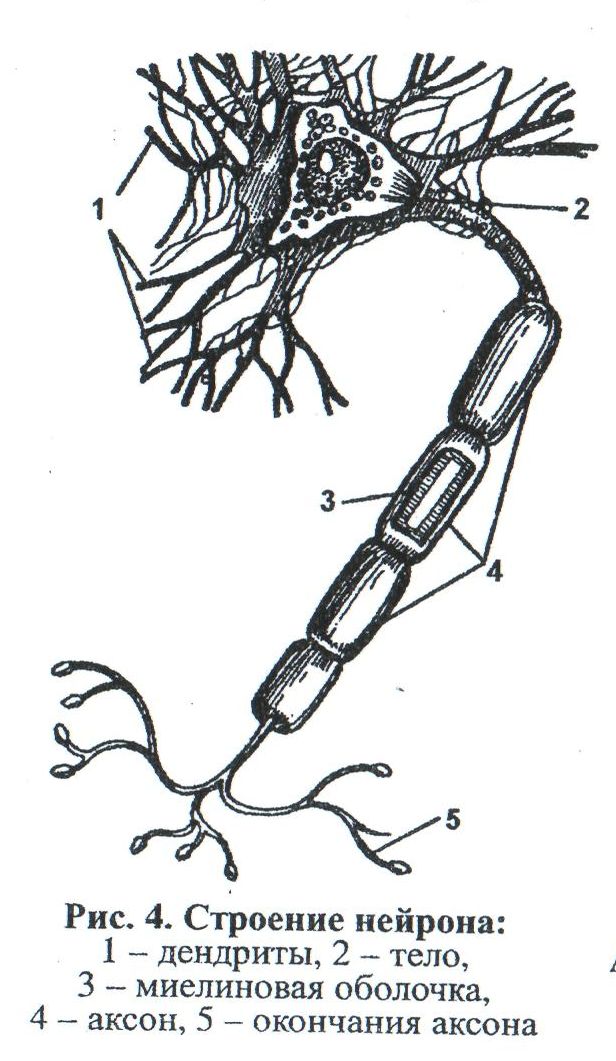
Nerve cells have a different shape and size and consist of a body and processes (Fig. 6).
The processes of the nerve cell are divided into two types:
- Neurites , or axons, along which excitation (impulse) is transmitted from the cell body to the periphery. The axon always departs alone from the cell and ends with the terminal apparatus in the working organ or on another neuron.
- Dendrites - processes along which an impulse is transmitted from the periphery to the cell body. There are many of them and they branch.
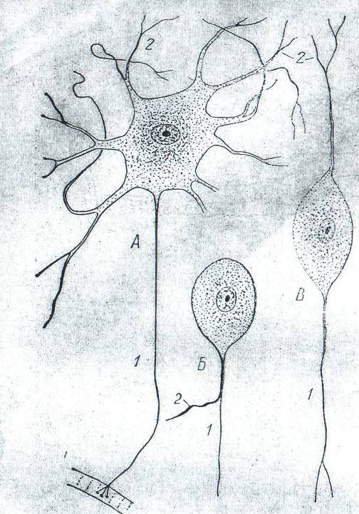
According to the number of processes, nerve cells are divided into three types (Fig. 7):
- Unipolar - cells with one branch. Not found in humans.
- Bipolar - have one neurite in the CNS and one dendrite going to the periphery. They are located in the spinal ganglions.
- Multipolar - have one neurite and many dendrites. A person has the most of them.
The nucleus of the nerve cell has a rounded shape and is located in the center.
In the cytoplasm of neurons there are neurofibrils, which are thin threads. In the body of the nerve cell, they form a dense network. In the processes, neurofibrils are arranged parallel to each other.
neuroglia represented by cells of various shapes with a large number of processes. There are more of these cells than nerve cells.
Nerve fibres.The processes of nerve cells with sheaths are called nerve fibers. Distinguish between myelinated (pulp) and non-myelinated (non-pulp). The processes are located in the center of the nerve fiber and are called the axial cylinder, which is covered with a sheath formed by neuroglial cells (lemmocytes).
unmyelinated fibers are an axial cylinder covered only by a sheath of lemmocytes.
myelinated - much thicker. They also consist of an axial cylinder, but they have two layers of membrane: an inner, thicker one - myelin, and an outer, thin one, consisting of lemmocytes. Outside, the myelin fiber is covered with a thin connective tissue sheath - neurilemma.
Nerve endings.All nerve fibers end in nerve endings. There are three groups:
- Efferent . They can be of two types: motor and secretory. Motor endings are the end devices of the axons of the somatic and autonomic nervous system.
- sensitive (receptors) are the terminal devices of the dendrites of sensitive neurons. They are divided into free, consisting of a branching of the axial cylinder, and non-free, containing all the components of the nerve fiber, covered with a capsule.
- end branches,forming interneuronal synapses, carrying out the connection of neurons with each other.
5. The concept of organs and organ systems
Connecting with each other, different tissues form organs.
An organ is a part of the body that has a certain shape, structure, occupies a certain place and performs a specific function. Various tissues take part in the formation of any organ, but only one of them is the main one, the rest perform an auxiliary function. For example, connective tissue forms the basis of an organ, epithelial tissue forms the mucous membranes of the respiratory and digestive organs, muscular tissue forms the walls of hollow organs (esophagus, intestines, etc.), nervous tissue is presented in the form of nerves innervating the organ and nerve nodes lying in the walls of organs. Organs differ in shape, size and position. In addition to individual differences, there are gender and age differences.
Organs that are similar in structure, origin and perform a single function are called a system. The following organ systems are distinguished in the human body:
- Musculoskeletal, which forms the skeleton of the body, ensures the movement of its parts in relation to each other and the movement of the organism in space.
- Respiratory , which determines the delivery of oxygen from the environment to the blood and the removal of CO from the body 2 as one of the end products of metabolism.
- The cardiovascular systemensures the movement of blood lymph through the blood and lymphatic vessels.
- Digestive systemdesigned for food processing, as well as for the absorption of nutrients into the blood and lymph.
- excretory systemensures the removal of metabolic products from the body.
- Endocrine , whose glands form hormones involved in the humoral regulation of body functions.
- reproductive system performs the function of reproduction and thereby maintains the existence of the species.
- Sensory system, perceiving irritations from the external world and the internal environment of the body.
- Nervous system regulates the state and activity of all systems.
References:
1. Textbook Loginov A.V. "Physiology with Fundamentals of Human Anatomy". M.: 1983.S.
Picture . The structure of various types of epithelium.
A - single-layer cylindrical; B - single-layer cubic; B - single-layer flat; G - multi-row; D - multilayer flat non-keratinizing; E - stratified squamous keratinizing; F 1 - transitional epithelium with a stretched wall of the organ and 2 - with a collapsed wall of the organ.
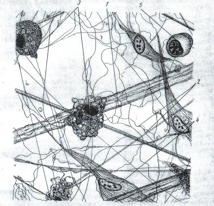
Drawing. Loose fibrous unformed connective tissue.
1-collagen fibers; 2- elastic fibers; 3- macrophages; fibroblasts; 5- plasma cell.
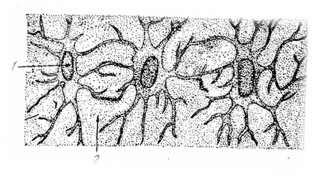
Picture. Bone cells.
1-processed bone cells; 2- intercellular substance.
Picture. Scheme of the structure of the osteon: in the left half, bone cavities and tubules are shown, in the right - the direction of the fibers in individual plates.
Picture. Types of muscle tissue.
Picture. The structure of a neuron.
1-dendrites; 2- body; 3- myelin sheath; 4 axon; 5- end of the axon.
Picture. Nerve cells.
A. multipolar neuron; B. unipolar neuron; B. bipolar neuron; 1-neurite; 2- dendrites