“Tissues, definition, classification. Epithelial, connective, muscle, nervous tissues
Methodical development
Topic: “Tissues, definition, classification.
epithelial, connective, muscular,
nervous tissue.
Speciality:
060108 "Pharmacy"
2008 - 2009 academic year
State educational institution SPO
"Orekhovo-Zuevsky Medical College"
Methodical development
discipline: "Human Anatomy and Physiology"
Section: "The body and its components"
Topic:
“Tissues, definition, classification. Epithelial, connective, muscle, nervous tissues.
Speciality:
060109 "Pharmacy"
Lecturer: Prikhodko E.I.
2008 - 2009 academic year
Class type:
Lecture lesson.
Location:
Medical College room №203 "Human Anatomy and Physiology"
Lesson duration:
Group:
Rationale for the choice of the topic of the lesson:
Topic: “Tissues, definition, classification. Epithelial, connective, muscle, nervous tissue" is included in the section "The body and its components" and meets the requirements of the state educational standard, which provides for the use of students' knowledge in practical activities and in the study of special disciplines. The topic is relevant, because on the basis of the knowledge gained, students will be able to further use the acquired knowledge when studying the course "Human Anatomy and Physiology".
Justification for choosing the type of lesson:
Given the large amount of material and the complexity of assimilation, the lecture session on the study and primary consolidation of new material has the form of explanatory and illustrative training using multimedia technologies (presentations).
Lesson objectives:
Didactic.
Formation of students' knowledge on the topic: “Tissues, definition, classification. Epithelial, connective, muscle, nervous tissues. Application of knowledge in the study of special disciplines and in practical activities.
Educational.
Formation of professionally significant qualities of a specialist's personality, instilling love for the chosen profession. Education in students of a conscientious attitude to study and work.
Developing.
Development of cognitive processes, students' abilities, development of logical thinking.
Occupational profile:
The student must submit the formation of various types of tissues from the germ layers, the location of different types of tissues and the function in the body.
Know:
Types of fabrics;
Differences between different types of fabrics;
The structure of epithelial, muscle, connective and nervous tissue.
Be able to:
Show on the tables the structure of epithelial, connective, nervous and muscle tissues;
Distinguish different types of fabrics on the tables.
Lesson equipment map.
Tables: "Epithelial tissue", "Connective tissue", "Blood", "Bone tissue", "Skeletal muscle tissue", "Cardiac muscle tissue", "Muscle fiber structure", "Nervous tissue". Graphs of the logical structure, atlases.
Methodical model of the lesson.
Organizational moment 5 minutes. Familiarization with the topic and lesson plan. Motivation of the theme of the lesson.
Control of the initial level of knowledge 10-15 minutes. front poll.
The main part of the lecture is 65-70 minutes. Lecture of explanatory and illustrative nature with the use of tables, with a phased consolidation.
Summing up 5 minutes. The logical conclusion of the lesson. Homework assignment.
2. "Human Anatomy" R.P. Samusev, Yu.M.Selin p.35-57
Literature for students:
1. "Anatomy and human physiology" E.A. Vorobiev, A.V. Gubar, E.B. Safyannikova pp. 4-8, 28-52.
Guidelines for teachers
by the stages of the lesson.
| No. p / p | Stages of the lesson and content | Methodological substantiation | Explanations for teachers |
| 1. | Organizing time:
Lesson plan:
| Organizes and disciplines students. Creates a working environment. | The teacher explains the importance of this topic for the study of special disciplines and the importance of knowledge for a pharmaceutical worker. |
| 2. | Control of the initial level of knowledge. Application No. 1 | front poll. Checking students' knowledge on the topic | Frontal survey |
| 3. | Learning new material:
5) Nervous tissue. Application No. 5. 6) Muscle tissue. Application No. 6. | Working with tables. Working with logical structure graphs. Working with tables and graphs of the logical structure Working with tables and graphs of the logical structure. Working with tables and graphs of the logical structure. | To draw students' attention to the structural features of various types of tissues. To draw students' attention to the formation of various types of tissues in embryonic development. To draw students' attention to the structural features of epithelial tissue. To draw students' attention to the structural features of the connective tissue. Draw students' attention to the structural features of the nervous tissue. Draw students' attention to the structural features of muscle tissue |
| 4. | Summing up the lesson. | Motivation for the next lesson, activation of self-training. |
Application No. 1.
Questions for the frontal survey.
Define the science of anatomy and physiology.
Define the term organ.
Name the organ systems.
Describe hollow and parenchymal organs.
Define an organ system.
List the planes of the human body.
List the axes of the human body, how they are formed.
List the basic anatomical concepts that determine the position of organs, their parts in the body.
Application number 2.
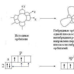
Textile- This is a system of cells and intercellular structures that have a common development, structure and perform a specific function.
Textile


Nervous Epithelial Connective Muscular
Application No. 3.
epithelial tissue covers the surface of the body and cavities of various tracts and ducts, with the exception of the heart, blood vessels and some cavities. In addition, almost all glandular cells are of epithelial origin. Layers of epithelial cells on the surface of the skin protect the body from infection and external damage. The cells that line the digestive tract from the mouth to the anus have several functions: they secrete digestive enzymes, mucus, and hormones; absorb water and food products. The epithelial cells that line the respiratory system secrete mucus and remove it from the lungs along with the dust and other foreign particles it traps. In the urinary system, epithelial cells carry out the excretion and reabsorption (reabsorption) of various substances in the kidneys, and also line the ducts through which urine is excreted from the body. Derivatives of epithelial cells are human germ cells - eggs and sperm, and the entire path that they pass from the ovaries or testes (genitourinary tract) is covered with special epithelial cells that secrete a number of substances necessary for the existence of an egg or sperm. The epithelium is a layer covering the internal and external surfaces of organisms. Its main function is to protect the relevant organs from mechanical damage and infection. In those places where the body tissue is subjected to constant stress and friction and "wears out", epithelial cells multiply at a high speed. Often, in places of heavy loads, the epithelium is compacted or keratinized. The free surface of the epithelium can also perform the functions of absorption, secretion and excretion, and perceive irritation. The epithelial cells are held together by a cementing substance containing hyaluronic acid. Since blood vessels do not approach the epithelium, the supply of oxygen and nutrients occurs by diffusion through the lymphatic system. Nerve endings can penetrate the epithelium.
Depending on the shape of the cell and the number of cell layers, the epithelium is divided into several types.
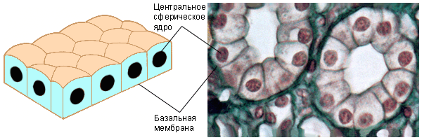
The least specialized of all is the cuboidal epithelium. Its cells, as the name implies, are cubic in cross section. This type of epithelium lines the ducts of many glands and also performs secretory functions within them.
cuboidal epithelium.
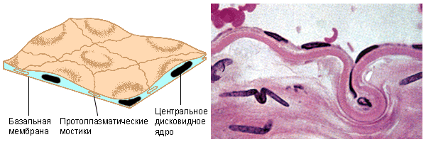
Squamous epithelial cells are thin and flattened; protoplasmic bonds they are tightly connected to each other. Due to this, they do not prevent the diffusion of various substances into the organs that these cells line: the alveoli of the lungs, the walls of the capillaries.
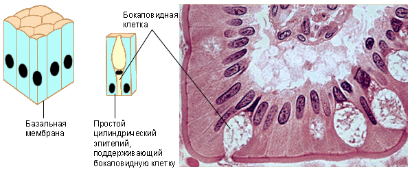
Tall and rather narrow cylindrical epithelial cells line the stomach and intestines. Scattered among cylindrical cells, goblet cells secrete mucus that protects these organs from self-digestion, and at the same time provide a lubricant to help move food. Microvilli are often found on the free surface of cells, increasing the suction surface.
flat epithelium.
cylindrical epithelium.
Ciliated epithelium.
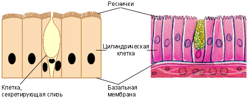
The ciliated epithelium is similar to the cylindrical, but carries numerous cilia on its surface. It lines the oviducts, the ventricles of the brain, the spinal canal, and the airways.
Stratified epithelium.
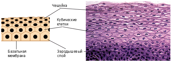
Stratified epithelium consists of several layers of cells; inside cubic, and outside - flatter, called scales. The thickness of this tissue is sufficient to protect the covered organs from leakage of various substances and mechanical damage. Scales can remain alive (for example, in the esophagus, gland ducts) or keratinize, turning into keratin (outer surface of the skin, buccal mucosa, vagina). The cells of the stratified transitional epithelium (bladder, ureter) are able to stretch.
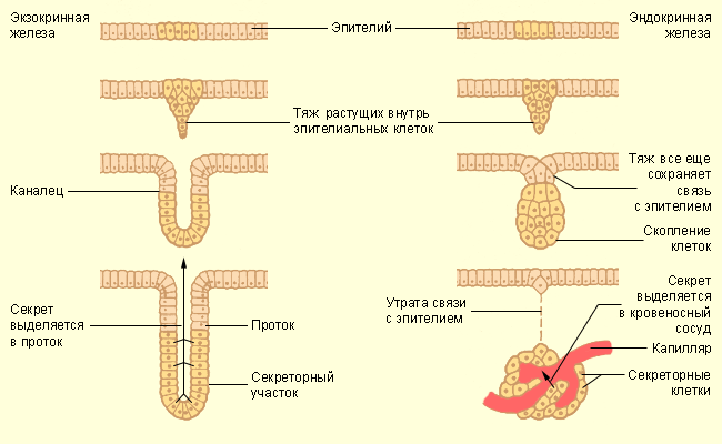
Sometimes goblet secretory cells form a multicellular gland. Exocrine glands secrete a secret to the surface of the epithelium, while endocrine glands are not connected with the epithelium and secrete a secret into the capillaries penetrating them. Products produced by the glands can be removed from the cell in three ways:
merocrine mechanism (sweat glands, etc.): secretion occurs through the membrane, and the cytoplasm is not consumed;
apocrine mechanism (mammary glands): together with the secret, the outer layers of the cytoplasm are rejected;
holocrine mechanism (sebaceous glands): the entire cell is destroyed.
 |
Application No. 4.
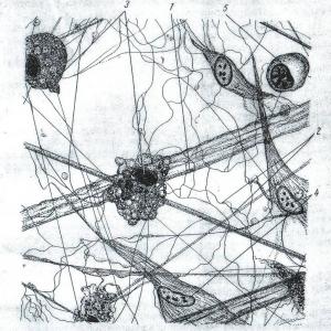
Connective tissue, or tissues of the internal environment, is represented by a group of tissues that are diverse in structure and functions, which are located inside the body and do not border on either the external environment or organ cavities. Connective tissue protects, insulates and supports parts of the body, and also performs a transport function within the body (blood). For example, the ribs protect the organs of the chest, the fat is an excellent insulator, the spine supports the head and torso, and the blood carries nutrients, gases, hormones, and waste products. In all cases, the connective tissue is characterized by a large amount of intercellular substance. The following subtypes of connective tissue are distinguished: loose, adipose, fibrous, elastic, lymphoid, cartilage, bone, and blood. Connective tissue is the main support of the animal body. It makes up the skeleton, connects various tissues and organs, surrounds some organs, protecting them from damage. Connective tissue consists of cells of various types, usually located far from each other; their oxygen and nutrient requirements are generally low.
Loose connective tissue consists of cells scattered in the intercellular substance and intertwined disordered fibers. Wavy bundles of fibers are made of collagen, while straight ones are made of elastin; their combination ensures the strength and elasticity of the connective tissue. On a transparent semi-liquid matrix containing these fibers, cells of various types are scattered:
oval mast cells surround blood vessels; they produce a matrix, and also produce heparin (anti-blood clotting) and hysparin (vasodilation, muscle contraction, stimulation of gastric juice secretion);
fibroblasts - cells that produce fibers;
macrophages (histocytes) - amoeboid cells that absorb pathogens;
plasma cells are another component of the immune system;
chromatophores - highly branched cells containing melanin; present in the eyes and skin;
fat cells;
mesenchymal cells - undifferentiated connective tissue cells that can, if necessary, turn into cells of one of the above types.
Dense connective tissue is made up of fibers, not cells. White tissue is found in tendons, ligaments, cornea, periosteum, and other organs. It consists of strong and flexible collagen fibers assembled in parallel bundles. Yellow connective tissue is found in ligaments, walls of arteries, and lungs. It is formed by a random weave of yellow elastic fibers.
Adipose tissue contains mainly fat cells. The fat cell consists of a central fat droplet, and the nucleus and cytoplasm are pushed back to the membrane. This type of tissue protects the underlying organs from shock and hypothermia.
Skeletal tissues are represented by cartilage and bone. Cartilage is a strong tissue consisting of cells (chondroblasts) immersed in an elastic substance - chondrin. Outside, it is covered with a denser perichondrium, in which new cartilage cells are formed. Cartilage covers the articular surfaces of bones, is found in the ear and pharynx, in articular bags and intervertebral discs.
The skeleton of vertebrates is built from bone. It consists of cells immersed in a solid substance consisting of 30% organic matter (mainly collagen) and 70% hydroxyaparite Ca 10 (PO 4) 6 (OH) 2 . It also contains sodium, magnesium, potassium, chlorine and other substances. This combination of materials greatly increases the resistance of bone tissue to stretching and bending. Bone cells (osteoblasts) are located inside special lacunae, interconnected by blood vessels.
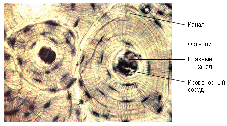 |
Bone tissue is divided into three types. Spongy bone is made up of thin bony elements called trabeculae; the space between them is filled with yellow (fat cells) or red (erythrocytes) bone marrow. On a section of dense bone tissue, one can see numerous cylinders formed by concentric bone plates. In the center of each such cylinder there is a Haversian canal through which an artery and a vein, a lymphatic vessel and nerve fibers pass. Membrane bone tissue does not have cartilaginous rudiments, but is formed directly in the skin layer. Spongy bone is characteristic mainly for embryos, and membranous bones are found in the skull, lower jaw and shoulder girdle.
Dentin is similar in composition to bone, but contains more inorganic matter. There are no gaps or haversian systems here. Dentin cells are located on its inner surface, blood vessels and nerve endings penetrating the tooth, as well as special processes that produce collagen, depart from them.
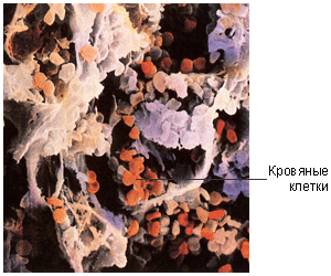
Myeloid tissue (bone marrow) produces blood cells - erythrocytes and granulocytes. Lymphoid tissue produces lymphocytes.
Blood cells in the bone marrow.
 |
Application No. 5
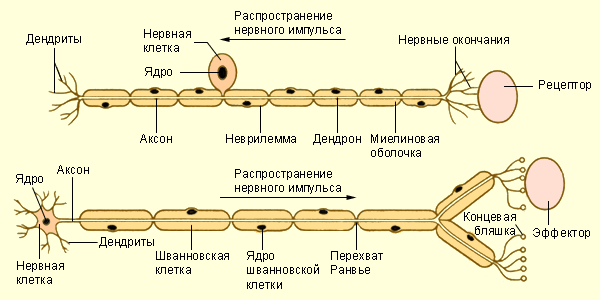
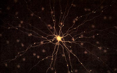
nervous tissue characterized by the maximum development of such properties as irritability and conductivity. Irritability - the ability to respond to physical (heat, cold, light, sound, touch) and chemical (taste, smell) stimuli (irritants). Conductivity - the ability to transmit an impulse (nerve impulse) that has arisen as a result of irritation. The element that perceives irritation and conducts a nerve impulse is a nerve cell (neuron). A neuron consists of a cell body containing a nucleus, and processes - dendrites and an axon. Each neuron may have many dendrites, but only one axon, which, however, has several branches. Dendrites, perceiving a stimulus from different parts of the brain or from the periphery, transmit a nerve impulse to the body of the neuron. From the cell body, a nerve impulse is conducted along a single process - an axon - to other neurons or effector organs. The axon of one cell can contact either dendrites, or the axon or bodies of other neurons, or muscle or glandular cells; these specialized contacts are called synapses. The axon extending from the cell body is covered with a sheath formed by specialized (Schwann) cells; the sheathed axon is called a nerve fiber. Bundles of nerve fibers make up nerves. They are covered with a common connective tissue sheath, in which elastic and non-elastic fibers and fibroblasts (loose connective tissue) are interspersed along the entire length. In the brain and spinal cord, there is another type of specialized cells - neuroglial cells. These are auxiliary cells contained in the brain in very large quantities. Their processes braid the nerve fibers and serve as a support for them, as well as, apparently, insulators. In addition, they have secretory, trophic and protective functions. Unlike neurons, neuroglial cells are capable of dividing. Nervous tissue consists of nerve cells - neurons and neuroglia cells. In addition, it contains receptor cells. Nerve cells can be excited and transmit electrical impulses.
Neurons consist of a cell body with a diameter of 3–100 µm, containing a nucleus and organelles, and cytoplasmic processes. Short processes that conduct impulses to the cell body are called dendrites; longer (up to several meters) and thin processes that conduct impulses from the cell body to other cells are called axons. Axons connect with neighboring neurons at synapses.
Different types of neurons.
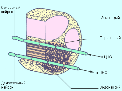 |
Neurons that transmit impulses to effectors (organs that respond to stimuli) are called motor neurons; neurons that transmit impulses to the central nervous system are called sensory. Sometimes sensory and motor neurons are interconnected using intercalary (intermediate) neurons.
 |
| The structure of sensory and motor nerves. |
Bundles of nerve fibers are assembled into nerves. The nerves are covered by a connective tissue sheath called the epineurium. Its own sheath covers each fiber individually. Like neurons, nerves are either sensory (afferent) or motor (efferent). There are also mixed nerves that transmit impulses in both directions. Nerve fibers are entirely or completely surrounded by Schwann cells. There are gaps between the myelin sheaths of Schwann cells called nodes of Ranvier.
Retinal neuron.
 |
Neuroglia cells are concentrated in the central nervous system, where their number is ten times greater than the number of neurons. They fill the space between neurons, providing them with nutrients. It is possible that neuroglial cells are involved in storing information in the form of RNA codes. When damaged, neuroglial cells actively divide, forming a scar at the site of damage; neuroglial cells of a different type turn into phagocytes and protect the body from viruses and bacteria.
Application No. 6
Muscle. Muscles provide movement of the body in space, its posture and contractile activity of internal organs. The ability to contract, to some extent inherent in all cells, is most strongly developed in muscle cells. There are three types of muscles: skeletal (striated, or voluntary), smooth (visceral, or involuntary), and cardiac.
Muscle tissue is made up of highly specialized contractile fibers. In organisms of higher animals, it is up to 40% of body weight.
Longitudinal sections of striated, smooth and cardiac muscle.
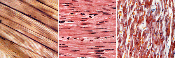 |
There are three types of muscles. Striated (also called skeletal) muscles are the basis of the body's motor system. Very long multinucleated fiber cells are connected to each other by a connective tissue containing many blood vessels. This type of muscle is distinguished by powerful and fast contractions; combined with a short refractory period, this leads to rapid fatigue. The activity of striated muscles is determined by the activity of the brain and spinal cord.
Smooth (involuntary) muscles form the walls of the respiratory tract, blood vessels, digestive and genitourinary systems. They are distinguished by relatively slow rhythmic contractions; activity depends on the autonomic nervous system. Mononuclear smooth muscle cells are collected in bundles or layers.
Finally, the cells of the heart muscle branch at the ends and are interconnected by means of superficial processes - intercalated discs. Cells contain several nuclei and a large number of large