Fabrics
The organs of the human body are made up of various tissues. Each tissue is a single system of cells and intercellular substance, which has a specific structure and performs a specific function in the body. The structure and function of the tissue developed in the course of the evolution of the animal world. When the conditions in which the body is located, in particular with various diseases, changes occur in the tissues. Depending on the structure and function, the following types of tissues are distinguished: epithelial, connective, muscle and nervous.
Epithelial tissue
Epithelial tissues make up the surface layer of the skin, line the mucous and serous membranes from the inside and form glands.
Common to all types of epithelial tissues is that they are built predominantly from cells; there is very little intercellular substance in them. Epithelial cells have different shapes and usually form sheets. The epithelium is separated from the underlying tissues by a thinnest plate called basement membrane.
Depending on the shape of the cells, there are three main types of epithelium: flat, cubic and cylindrical(fig. 3). In this case, the cells can be located in one layer - unilamellar epithelium and in several layers - stratified epithelium... In a stratified epithelium, the cells of each layer usually have their own characteristics (shape, size, etc.). The functions of the epithelium include a protective metabolism between the body and the external environment, etc.
In accordance with the structural features and functional properties, the following main types of epithelial tissue are distinguished.
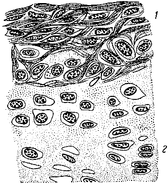
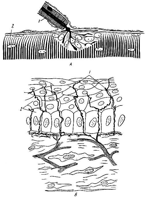
Stratified squamous epithelium forms the surface layer of the skin and some mucous membranes of the oral cavity, pharynx, urinary organs (renal pelvis, ureters, bladder, urethra), etc. The skin epithelium consists of many tens of layers of cells that differ in shape and function. The deepest layer is called sprout... It is formed by cylindrical cells, due to which the restoration of other epithelial cells occurs. Next are the layers of spiny cells connected by their processes - thorns - to each other. Outside, the cells flatten, and the superficial layers of the epithelium consist of thin plates that contain dense stratum corneum and gradually fall off. The epithelium of the skin performs a protective function: it protects the body from various chemical, thermal and mechanical influences. At the same time, it also participates in the metabolism: through it, some decay products are released and heat transfer.
The stratified squamous epithelium of the urinary tract mucosa is called the transitional epithelium, since it changes its thickness and structure depending on the filling of the organ: when the organ wall contracts, the thickness of the epithelium increases, and when stretched, it decreases; the shape of the cells also changes somewhat.
Monolayer columnar or prismatic epithelium lines the mucous membrane of the stomach, small and large intestine and some other organs. As the name suggests, this epithelium consists of a single layer of cells. It plays a protective role, protecting the underlying tissues from the digestive action of digestive juices. The cells of the epithelium of the small intestine have a special formation on their surface - a border, which consists of many microvilli that facilitate the absorption of nutrients.
Unilamellar ciliated epithelium lining the mucous membrane of the respiratory tract, consists of cells of various shapes, on the surface of which there are ciliated cilia. Wave-like vibrating in the direction opposite to the stream of inhaled air, the cilia expel dust particles settling from the air onto the mucous membrane. The ciliated epithelium of the airways plays mainly a protective role. In humans, ciliated epithelium is also present in the fallopian tubes; here, the vibrations of the cilia promote the movement of the egg.
Unilamellar cubic epithelium lines the urinary tubules of the kidneys and participates in the process of urine formation. Cubic epithelium is also found in the small excretory ducts of many glands and in the small bronchi (here it is provided with cilia).
Monolayer squamous epithelium, or mesothelium, lines the membranes of the internal body cavities - the serous membranes (peritoneum, pleura and pericardium). Covering the sheets of serous membranes facing each other, the mesothelium prevents the organs covered with serous membranes from growing together with each other. In addition, the mesothelium is involved in the formation and absorption of serous fluid. Serous fluid is in the form of a thin layer between the layers of the serous membrane, which reduces friction when they are displaced.
Glandular epithelium makes up the main tissue of special organs - glands. The cells of the glandular epithelium have the ability to form and secrete special substances. This function of the glands is called secretory, and the substances secreted by the glands are called secrets... In some cases, the ability to secrete secretions is possessed by individual cells that are part of the epithelial layer; this is - unicellular glands(for example, goblet cells of the intestine that secrete mucus).
In other cases, specific secretions are secreted by complex organs - multicellular glands. Such glands are salivary glands, thyroid gland, etc. Some glands have excretory ducts and are called excretory glands, other glands do not have excretory ducts, secrete their secretions directly into the blood and are called endocrine glands.
Connective tissue
Connective tissues are composed of cells and intercellular substance. Unlike other tissues, the intercellular substance in the connective tissues is expressed as well as the cells; it is represented by various fibers and a basic amorphous substance. Depending on the features of the structure and function, several types of this group of tissues are distinguished: loose fibrous connective tissue, adipose tissue, reticular tissue, dense fibrous connective tissue, cartilaginous tissue, bone tissue, etc. The functions of various types of connective tissue are as follows: trophic (trophic - nutrition ), supporting and protective.
The group of connective tissues usually includes blood and lymph. The structure of blood and lymph and their functions are described in chapter X.
Loose fibrous connective tissue(Fig. 4) is widely distributed in the body. It accompanies blood vessels, forms the skeleton of many organs and layers between organs, is part of the subcutaneous layer, etc. The main cells of this tissue are macrophages, fibroblasts, adventitia, etc. Macrophages are capable of amoeboid movement and phagocytosis, i.e., active seizure bacteria and other particles and their digestion (if these particles are organic). Phagocytosis is a protective reaction of the body. The phenomenon of phagocytosis was first described by the Russian scientist I.I.Mechnikov.
![]()
Fibroblasts are involved in the formation of intercellular substance, in particular connective tissue fibers; adventitia cells can transform into other cell forms.
The intercellular substance of loose fibrous connective tissue is formed by a basic, structureless (amorphous) viscous substance and various fibers lying in it. Collagen(or adhesive) fibers are thin, unbranched, form bundles and have little elasticity. Elastic fibers are thin, branching, do not form bundles; they stretch easily and, after the elimination of the force causing them to stretch, quickly return to their previous state.
Loose fibrous connective tissue performs a supporting, protective and trophic function in the body. Support function carried out due to its fibers, which create the stroma (base) of the organ, give it strength and elasticity. Protective function performed by macrophages - cells that actively participate in the fight against microbes entering the body - pathogens. Trophic function- this is participation in the process of nutrition of tissues of various organs. This role in loose fibrous connective tissue is played by its main substance. Nutrients enter the tissues of organs from the blood through the walls of blood vessels, and the latter is always accompanied by connective tissue. Thus, in order to get to all tissues of the organ, nutrients must pass through the walls of blood vessels and the adjacent connective tissue. What substances, in what quantity and at what speed will pass into the organs - depends on the state of the vascular wall and the main substance of the connective tissue.
Adipose tissue is a type of loose fibrous tissue, constitutes the subcutaneous tissue, the layers around the vessels and many organs, is part of the omentum, etc. This tissue, along with cells and intercellular substance inherent in loose fibrous connective tissue, contains a large number of fat cells. Adipose tissue performs mainly a trophic function, since it contains a reserve of fat, which, if necessary, is consumed by the body. Fat layers also perform a mechanical function, protecting some organs (for example, blood vessels) from damage.
Reticular tissue is the basis of the hematopoietic organs and is part of some other organs. In this tissue, the cells are interconnected with the help of processes of the cytoplasm. Such structures are called syncytium. Cells capable of phagocytosis, macrophages, can be separated from the syncytium. Like loose connective tissue, the reticular tissue performs a trophic and protective function, the supporting role of this tissue is insignificant.
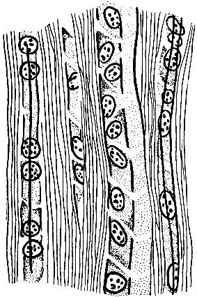
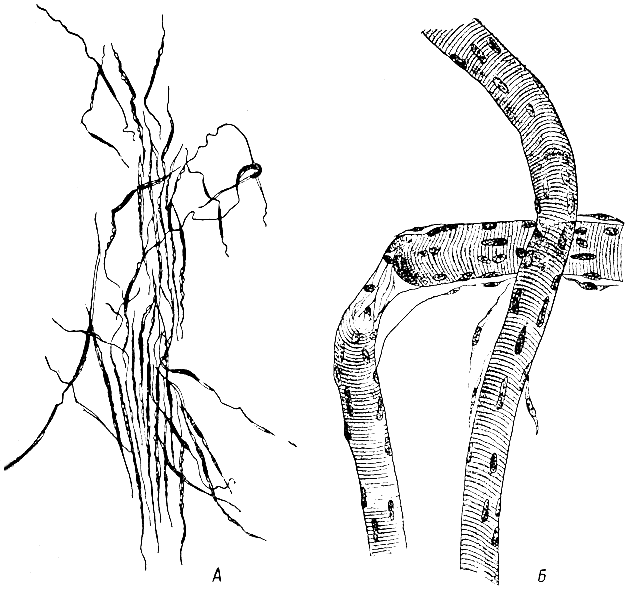
Dense fibrous connective tissue forms tendons (Fig. 5), ligaments, the basis of the skin (the skin itself) and performs a supporting function. This type of tissue is distinguished by a highly developed intercellular substance. Bundles of collagen fibers achieve especially powerful development; there are also elastic fibers. There is little structureless substance. Cells are located between the fibers: fibrocytes, etc.
Cartilage tissue... Depending on the structure of the intercellular substance, three types of cartilage tissue are distinguished: hyaline, elastic and fibrous cartilage. Cartilage of all kinds has a mechanical function.
From hyaline cartilage(Fig. 6) the cartilaginous parts of the ribs, most of the cartilage of the larynx and the articular cartilages of most joints are formed. Under the microscope, the intercellular substance of the hyaline cartilage appears as a vitreous homogeneous mass. However, using special methods, it is possible to find out that it consists of a basic structureless substance and fibers similar in structure to collagen fibers. The main substance contains cartilage cells in oval-shaped capsules.
Elastic cartilage forms the base of the auricle and epiglottis. It differs from hyaline cartilage in that it has a dense network of elastic fibers in the base substance.
Fibrous, or connective tissue, cartilage occurs in some joints of bones (for example, in the intervertebral discs) and in the places where tendons attach to bones. In the intercellular substance of this cartilage there is a large number of parallel, well-defined bundles of collagen fibers; there is very little basic substance.
All types of cartilage from the surface are covered perichondrium, which is a type of dense fibrous connective tissue. From the side of the perichondrium, the cartilage is nourished and grown.
Bone... Bone tissue is represented by bone cells - osteocytes - and intercellular substance (Fig. 7). Osteocytes are cells whose processes are connected to each other. The cell bodies are located in special bone cavities, and their processes are in the so-called bone tubules. The intercellular substance is built from the main structureless substance and fibers, similar in composition and properties to collagen. However, unlike other types of connective tissue, the intercellular substance of bone tissue contains mineral salts (calcium phosphate, calcium fluoride, etc.), which give it special strength.
The basic structural unit of bone is osteon(Fig. 8), which is a system of concentrically located bone plates. They are in the form of cylinders inserted into each other and are called Havers plates. In the center of the osteon there is a canal called the Haversian channel. The Haversian canals contain blood vessels that originate from larger vessels entering the bone through the so-called nutrient canals. Intercalated bone plates are located between the osteons.
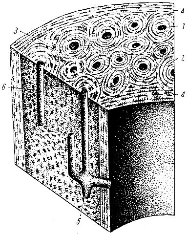
There are also external and internal common bone plates.
Muscle tissue
This group includes tissues of various structures and origins: smooth muscle tissue and striated muscle tissue. What they have in common is the ability to contract.
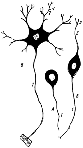
Smooth muscle tissue... Smooth muscle tissue is part of the walls of internal organs (intestines, bladder, uterus, etc.) and blood vessels, and is located in the skin. Its structural element is muscle fiber. This is a spindle-shaped cell (Fig. 9, A) with a length of 60 - 100 μ; it consists of sarcoplasm (i.e., cytoplasm), inside which is a rod-shaped nucleus. In the sarcoplasm there are special structures - contractile filaments or myofibrils.
Striated muscle tissue... Striated muscle tissue (Fig. 9, B) is located in skeletal muscles and in some internal organs (tongue, soft palate, etc.). Its structural element is muscle fibers, the length of which in humans can reach 12 cm with a diameter of 2 to 70 μ. Each muscle fiber, in addition to sarcoplasm, contains a large number of nuclei and has a membrane. In the contractile filaments (myofibrils) of striated muscle fibers under a microscope, alternating dark and light areas can be distinguished, which give these fibers a transverse striation (hence the name of the tissue). Dark and light discs of myofibrils have different physicochemical properties, in particular, the effect of birefringence is observed in dark discs, this effect is absent in light discs. Using an electron microscope, it was found that myofibrils, in turn, consist of even thinner filaments - protein filaments, or protofibrils, consisting of muscle protein molecules. Distinguish between thin and thick protofibrils. Muscle fibers form bundles separated from each other by layers of loose connective tissue.
Nerve tissue
Nerve tissue- the main element of the nervous system, which regulates the processes occurring in the body, and realizes its relationship with the environment.
The main properties of nervous tissue are excitability and conductivity. In response to various stimuli acting on the body from the external environment, or to changes occurring in the body itself, excitation (nerve impulses) arises in the nervous system.
Nerve tissue is formed by nerve cells and neuroglia.
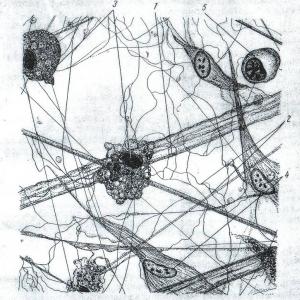
Nerve cell, or neuron(Fig. 10), consists of a cell body and its processes. According to the number of processes, unipolar neurons are distinguished - with one process, bipolar - with two and multipolar - with three or more processes. There are also pseudo-unipolar cells; one process departs from the body of such a cell, which soon divides into two. Sensitive cells, communication cells (intercalary) and motor nerve cells are distinguished by functional characteristics. Each neuron has one (or more, depending on the type of neuron) processes along which the excitation is conducted to the body of the nerve cell - a dendrite, and one process along which the excitation is conducted from the body of the nerve cell - neuritis, or axon. Dendrites are usually short and branching, neuritis - Long. Only a few nerve cells have long dendrites.
In the body of a neuron, a nucleus and cytoplasm are distinguished - neuroplasm. In addition to the organelles usual for all cells (mesh apparatus, etc.), the cytoplasm of the neuron contains special formations associated with the specific function of the nervous tissue. These are neurofibrils, the thinnest filaments that, without interruption, pass through the cell body from one process to another. Another special structure of neuroplasm is the so-called tigroid substance (Nissl's substance); with the help of special methods, it is revealed in the form of grains and lumps and corresponds to the ergastoplasm of other cells. With prolonged work of the organ, which is innervated by the cell, the tigroid substance disappears, and at rest appears again.
Nerve fibers- processes of nerve cells - represent the cytoplasm with neurofibrils running through it. However, the shells of the processes are built differently. Depending on the structure of the shell, pulpy and non-pulpy nerve fibers are distinguished. The fleshy nerve fibers are provided with a sheath of a fat-like substance - myelin, the non-fleshy ones are devoid of this sheath. Nerve fibers have endings (Fig. 11), which are functionally subdivided into endings that perceive irritation, and endings that transmit excitation to the working organs. The first of them are called sensitive (receptors), the second - motor in the muscles and secretory in the glands (effectors). The switching of a nerve impulse from one cell to another occurs with the help of special devices - synapses. A synapse is the contact of two neurons, which ensure the transition of nerve excitation from one nerve cell to another.
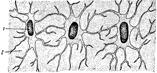
The second element of the nervous system is neuroglia. It is represented by cells of various shapes (Fig. 12), mainly dendritic (stellate and tree-branching). Neuroglial cells are present not only in the brain and spinal cord, but also accompany, in the form of the so-called Schwann sheath, nerve fibers leaving the brain.
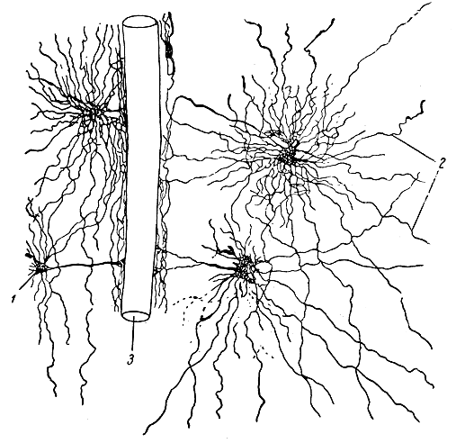
Neuroglia performs a trophic, protective and partially supporting function in the nervous tissue.