The structure of human organs
Organization levels
Man is the pinnacle of the evolution of the animal world. All living bodies are composed of separate molecules, which, in turn, are organized in cells, cells - in fabrics, fabrics - in organs, organs - in organ systems... And they together form a holistic organism.
The diagram shows the interconnection of all organ systems in the body. The determining (determinant) beginning is the genotype, and the general regulatory systems are the nervous and endocrine ones. The levels of organization from molecular to systemic are characteristic of all organs. The organism as a whole is a single interconnected system.
Life on Earth is represented by individuals of a certain structure, belonging to certain systematic groups, as well as to communities of varying complexity. Individuals and communities are organized in space and time. According to the approach to their study, several main levels of organization of living matter can be distinguished:
Molecular- any living system, no matter how complex it is organized, manifests itself at the level of functioning of biological macromolecules: nucleic acids, proteins, polysaccharides and other organic ones. The most important life processes begin from this level: metabolism and energy conversion, transmission of hereditary information, etc. This level is studied by molecular biology.
Cellular- a cell is a structural, functional and universal unit of a living organism. Cell biology (the science of cytology) studies the morphological organization of the cell, the specialization of cells during development, the function of the cell membrane, the mechanism and regulation of cell division;
Fabric- a set of cells united by a common origin, similarity in structure and the performance of a common function.
Organ- structural and functional association and interaction of several types of tissues that form organs.
Organic- an integral differentiated system of organs that perform various functions and represent a multicellular organism.
Population-specific- a set of individuals of the same species, united by a common habitat, creating a population as a system of supra-organismal order. In this system, the simplest elementary evolutionary transformations are carried out.
Biogeocenotic- a set of organisms of different types and varying complexity of organization with all factors of the environment.
Biosphere- a system of the highest rank, covering all the phenomena of life on Earth. At this level, the circulation of substances and the transformation of energy are carried out, associated with the vital activity of living organisms.
| Level | Structures | Functioning |
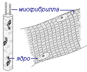
| Molecular | Proteins: actin, myosin | Energy release, movement of actin filaments relative to myosin filaments |
| Subcellular | Sarcomeres and myofibrils - structures formed by several proteins | Shortening of sarcomeres and myofibrils |
| Cellular | Muscle fibers | Shortening of muscle fibers |
| Tissue | Striated skeletal muscle tissue | Shortening of groups (bundles) of muscle fibers |
| Organic | Striated skeletal muscle | Muscle shortening |
| Systemic | Musculoskeletal system | Changing the position of bones (skin in the case of facial muscles) relative to each other |
| Functional system | Musculoskeletal system | Moving body parts or body in space |
Body structure
The senses are located on the head: unpaired - nose, tongue; paired - eyes, ears, organ of balance... Inside the skull is brain.
The human body is covered with skin. Bones and muscles form the musculoskeletal system. Inside the body are two body cavities - abdominal and thoracic separated by a septum - muscular diaphragm... In these cavities are located internal organs... In the chest - lungs, heart, blood vessels, airways and esophagus... In the abdominal cavity on the left (under the diaphragm) - stomach, on right - liver with gallbladder and spleen... In the canal of the spine there is spinal cord... In the lumbar region there are kidneys from which depart ureters included in bladder with urethra.
The female genitals are represented by: ovaries, fallopian tubes, uterus.
The male genitals are represented: testicles located in scrotum.
Organs and organ systems
Each organ has its own shape and a specific place in the human body. Organs performing general physiological functions are combined into an organ system.
| Organ system | System functions | Bodies that make up the system |
| Covering | Protecting the body from damage and the penetration of pathogens into it | Leather |
| Musculoskeletal | Giving strength and shape to the body, performing movements | Skeleton, muscles |
| Respiratory | Providing gas exchange | Respiratory tract, lungs, respiratory muscles |
| Circulatory | Transport, supply of all organs with nutrients, oxygen, excretion of metabolic products | Heart, blood vessels |
| Digestive | Digestion of food, providing the body with energy substances, protective | Salivary glands, teeth, tongue, esophagus, stomach, intestines, liver, pancreas |
| Excretory | Excretion of metabolic products, osmoregulation | Kidneys, bladder, ureters |
| Reproductive system | Reproduction of organisms | Ovaries, oviducts, uterus, testes, external genitalia |
| Nervous system | Regulation of the activity of all organs and body behavior | Brain and spinal cord, peripheral nerves |
| Endocrine system | Hormonal regulation of the work of internal organs and body behavior | Thyroid gland, adrenal glands, pituitary gland, etc. |
The nervous system regulates with the help of electrochemical signals, nerve impulses. The endocrine system works with the help of biologically active substances - hormones that enter the bloodstream and, reaching the organs, change their work.
Cellular structure of the body
External and internal environment of the body
External environment- this is the environment in which the human body is located. This is a set of specific abiotic and biotic conditions in which a given individual, population or species lives. A person lives in a gaseous environment.
The internal environment of the body is called the environment that is inside the body: it is separated from the external environment by the membranes of the body (skin, mucous membranes). It contains all the cells of the body. It is liquid, has a certain salt composition and a constant temperature. The internal environment does not include: the contents of the alimentary canal, urinary tract and respiratory tract. They border on the external environment: the outer stratum corneum and some mucous membranes. The organs of the human body supply the cells with the necessary substances through the internal environment and remove unnecessary substances during the life of the body.
Cell structure
Cells are diverse in shape, structure and functions, but similar in structure. Each cell is separated from others by a cell membrane. Most cells have a cytoplasm and a nucleus. Cytoplasm- the internal environment, the living contents of the cell, consisting of a fibrous basic substance - the cytosol and cellular organelles. Cytosol- the soluble part of the cytoplasm that fills the space between cellular organelles. Cytosol contains 90% water, as well as mineral and organic substances (gases, ions, sugars, vitamins, amino acids, fatty acids, proteins, lipids, nucleic acids, and others). This is the site of metabolic processes (for example, glycolysis, synthesis of fatty acids, nucleotides, amino acids, etc.).
In the cytoplasm of the cell there is a number of organelle structures, each of which has a specific function and has regular features of the structure and behavior at different periods of the cell's life. Organelles- permanent, vital constituents of cells.
Kernel structure and functions
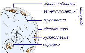
The cell and its contents are separated from the external environment or from neighboring cells by a surface structure. Core- the most important, obligatory organoid of the animal cell. It has a spherical or ovoid shape, 10–20 µm in diameter. The nucleus is separated from the cytoplasm by the nuclear membrane. The outer nuclear membrane from the surface facing the cytoplasm is covered with ribosomes, the inner membrane is smooth. The outgrowths of the outer nuclear membrane are connected to the channels of the endoplasmic reticulum. The exchange of substances between the nucleus and the cytoplasm is carried out in two main ways: through the nuclear pores and as a result of the lacing of the protrusions and outgrowths of the nuclear envelope.
The nuclear cavity is filled with gelatinous nuclear juice (karyoplasm), which contains one or more nucleoli, chromosomes, DNA, RNA, enzymes, ribosomal and structural proteins of chromosomes, nucleotides, amino acids, carbohydrates, mineral salts, ions, as well as products of the activity of the nucleolus and chromatin. Nuclear juice performs binding, transport and regulatory functions.
The cell nucleus, as the most important component of the cell, containing DNA (genes), performs the following functions:
- Storage, reproduction and transmission of hereditary genetic information.
- Regulation of metabolic processes, biosynthesis of substances, division, cell vital activity.
The nucleus contains chromosomes, the basis of which is DNA molecules that determine the hereditary apparatus of the cell. The regions of DNA molecules responsible for the synthesis of a particular protein are called genes... Each chromosome contains billions of genes. By controlling the formation of proteins, genes control the entire chain of complex biochemical reactions in the body and thereby determine its characteristics. Ordinary cells (somatic) of the human body contain 46 chromosomes, in sex cells (eggs and sperm) 23 chromosomes (half set).
In the core is nucleolus- a dense rounded body immersed in nuclear juice in which the synthesis of important substances is carried out. It is the center of the synthesis and organization of ribonucleoproteins, which in the form of bundles of filamentous formations form the chromatin structures of the nucleolus. Thus, the nucleolus is the site of RNA synthesis.
Cell organelles
Permanent cellular structures, each of which performs its own special functions, are called organelles... In the cell, they play the same role as organs in the body.
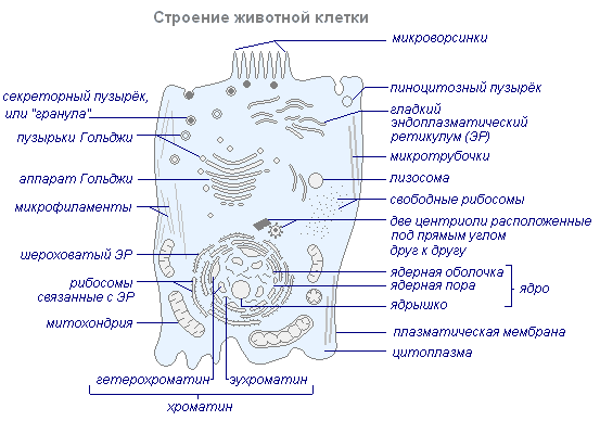
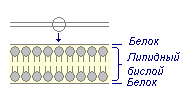
The main membrane structures of the cell are cytoplasmic membrane separating a cell from neighboring cells or intercellular substance, endoplasmic reticulum, Golgi apparatus, mitochondrial and nuclear membranes. Each of these membranes has structural features and specific functions, but they are all built according to the same type.
Functions cytoplasmic membrane:
- Restriction of the content of the cytoplasm from the external environment by the formation of a cell surface.
- Damage protection.
- Distribution of the intracellular environment into compartments in which certain metabolic processes take place.
- Selective transport of substances (semi-permeability). The outer cytoplasmic membrane is easily permeable to some substances and impermeable to others. For example, the concentration of K + ions is always higher in the cell than in the environment. On the contrary, there are always more Na + ions in the extracellular fluid. The membrane regulates the entry of certain ions and molecules into the cell and the excretion of substances from the cell.
- The energy transforming function is the conversion of electrical energy into chemical energy.
- Reception (binding) and transmission of regulatory signals into the cell.
- Secretion of substances.
- Formation of intercellular contacts, connection of cells and tissues.
Endoplasmic reticulum- a branched membrane system of channels with a diameter of 25–75 nm and cavities that penetrate the cytoplasm. There are especially many channels in cells with intensive metabolism, through which substances synthesized on membranes are transported.
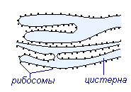
There are two types of membranes of the endoplasmic reticulum: smooth and rough(or granular containing ribosomes). On smooth membranes are enzyme systems involved in fat and carbohydrate metabolism, detoxification of substances. Such membranes predominate in the cells of the sebaceous glands, where fats are synthesized, liver (glycogen synthesis). The main function of rough membranes is protein synthesis, which is carried out in ribosomes. There are especially many rough membranes in glandular and nerve cells.
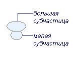
Ribosomes- small spherical bodies 15–35 nm in diameter, consisting of two subunits (large and small). Ribosomes contain proteins and r-RNA. Ribosomal RNA (r-RNA) is synthesized in the nucleus on the DNA molecule of some chromosomes. Ribosomes are also formed there, which then leave the nucleus. In the cytoplasm, ribosomes can be located freely or be attached to the outer surface of the membranes of the endoplasmic reticulum (rough membranes). Depending on the type of the synthesized protein, ribosomes can "work" singly or combine into complexes - polyribosomes. In such a complex, the ribosomes are linked by a long mRNA molecule. The function of ribosomes is participation in protein synthesis.
Golgi apparatus- a system of membrane tubes forming a stack of flattened sacs (cisterns) and associated systems of bubbles and cavities. The Golgi apparatus is especially developed in cells that produce protein secretions, in neurons, eggs. The tanks are connected by EPS channels. Proteins, polysaccharides and fats synthesized on EPS membranes are transported to the Golgi apparatus, condense inside its structures and "packaged" in the form of a secret, ready either for isolation or for use in the cell itself during its life. The Golgi apparatus is involved in the renewal of biomembranes and the formation of lysosomes.
Lysosomes- small rounded bodies, about 0.2-0.5 µm in diameter, bounded by a membrane. Inside the ribosomes there is an acidic environment (pH 5) and contains a complex (more than 30 types) of hydrolytic enzymes for the breakdown of proteins, lipids, carbohydrates, nucleic acids and others. There are several tens of lysosomes in a cell (there are especially many of them in leukocytes).
Lysosomes are formed either from the structures of the Golgi complex, or directly from the endoplasmic reticulum. They approach pinocytic or phagocytic vacuoles and pour their contents into their cavity. The main function of lysosomes is participation in the intracellular digestion of nutrients through phagocytosis and the secretion of digestive enzymes. Lysosomes can also break down and remove dead organelles and waste substances, destroy the structures of the cell itself during its death, during embryonic development and in a number of other cases.
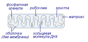
Mitochondria- small bodies, limited by a two-layer membrane. Mitochondria can have various shapes - spherical, oval, cylindrical, filamentous, spiral, elongated, cupped, branched. Their dimensions are 0.25–1 µm in diameter and 1.5–10 µm in length. The number of mitochondria in a cell is several thousand, in different tissues it is not the same, which depends on the functional activity of the cell: there are more of them where synthetic processes are more intense (for example, in the liver).
The mitochondrial wall consists of two membranes - an outer smooth and an inner folded one, into which an electron transport chain, ATPase, is built, and an intermembrane space 10–20 nm in size. Partitions extend from the inner membrane deep into the organoid, or crista... Folding significantly increases the inner surface of the mitochondria.
Numerous enzymes involved in energy metabolism (enzymes of the Krebs cycle, fatty acid oxidation, and others) are located on the membranes of the cristae in the mitochondrial matrix (inside the mitochondria). Mitochondria are closely associated with the membranes of the EPS, the channels of which often open directly into the mitochondria. The number of mitochondria can rapidly increase by division, which is due to the DNA molecule that is part of them. So, inside mitochondria contain their own DNA, RNA, ribosomes, proteins. The main function of mitochondria is the synthesis of ATP during oxidative phosphorylation (aerobic cell respiration).
| Schematic representation | Structure | Functions |
| Plasma membrane (cell membrane)
| Two layers of lipid (bilayer) between two layers of protein | Selectively permeable barrier that regulates the exchange between the cell and the environment |
| Core
| The largest organelle, enclosed in a membrane of two membranes, permeated with nuclear pores. Contains chromatin- in this form, untwisted chromosomes are in interphase. Contains nucleolus | Chromosomes contain DNA - the substance of heredity. DNA consists of genes regulating all types of cellular activity. Nuclear division is the basis of cell multiplication, and therefore the reproduction process. R-RNA and ribosomes are formed in the nucleolus |
| Endoplasmic reticulum (EPS)
| A system of flattened membrane sacs - cisterns - in the form of tubes and plates. Forms a single unit with the outer membrane of the nuclear envelope | If the surface of the EPS is covered with ribosomes, then it is called rough... Protein synthesized on ribosomes is transported along the cysterms of the EPS. Smooth(without ribosomes) serves as a site for the synthesis of lipids and steroids |
| Ribosome
| Very small organelles, consisting of two subparticles - large and small. They contain protein and RNA in approximately equal proportions. Ribosomes found in mitochondria are even smaller | The site of protein synthesis, where various interacting molecules are held in the correct position. Ribosomes are associated with EPS or lie freely in the cytoplasm. Many ribosomes can form a polysome (polyribosome) in which they are strung on a single strand of messenger RNA |
| Mitochondria
| The mitochondrion is surrounded by a membrane of two membranes; internal the membrane forms folds (cristae). Contains a matrix containing a small number of ribosomes, one circular DNA molecule and phosphate granules | During aerobic respiration, oxidative phosphorylation and electron transfer occurs in the cristae, and enzymes involved in the Krebs cycle and fatty acid oxidation work in the matrix. |
| Golgi apparatus
| A stack of flattened membrane sacs - cisterns. At one end of the stack, the pouches are continuously formed, and at the other, they are laced in the form of bubbles. | Many cellular materials (for example, EPS enzymes) undergo modification in cisterns and are transported in vesicles. The Golgi apparatus is involved in the process of secretion, and lysosomes are formed in it. |
| Lysosome
| Simple spherical membrane pouch (single membrane) filled with digestive (hydrolytic) enzymes | It performs many functions, always associated with the disintegration of any structures or molecules. Lysosomes play a role in autophagy, autolysis, endocytosis, exocytosis |


Cell division
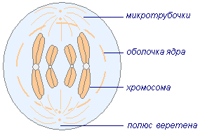
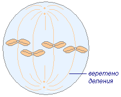
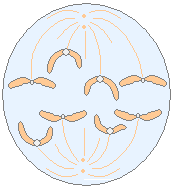
Cell division is a complex process of asexual reproduction. In unicellular organisms, it leads to an increase in the number of individuals, while multicellular organisms that begin their existence from one cell - zygotes, create a multicellular organism. This is a complex process, starting with the formation of the same molecule next to each DNA molecule. Thus, there are two identical DNA molecules in the chromosome. Before the start of cell division, the nucleus increases in size. The chromosomes are twisted into a spiral, and the nuclear envelope disappears. Organelles of the cell center diverge to opposite poles and between them is formed spindle division. Then the chromosomes line up along the equator. Paired DNA molecules of each chromosome are linked to centrioles- one DNA molecule with one centriole, and its double with the other. Soon, DNA molecules begin to diverge (each to its own pole), forming new sets of identical chromosomes and genes. In daughter cells, chromosomal tangles are formed, around which a nuclear envelope is formed. The chromosomes unwind and are no longer visible. After the nucleus is formed, the organelles divide, the cytoplasm - a constriction appears dividing one cell into two daughter cells.
| Fission phases | Drawing | Mitosis |
| Prophase |
|
|
| Metaphase |
|
|
| Anaphase |
|
|
| Telophase |
|
|
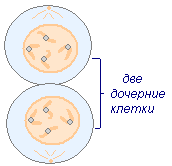
The biological significance of mitosis consists in reproducing an identical cell, maintaining a constant number of chromosomes. The result of his work is the formation of two genetically homogeneous cells identical to the mother.
Cell life processes
Processes are going on in the cells of any organism metabolism... Nutrients entering the cell form complex substances; cellular structures are formed. In addition, with the formation of new substances, the processes of biological oxidation of organic substances - carbohydrates, proteins, fats - take place, while the energy necessary for the life of the cell is released, and the decay products are removed.
Enzymes... The synthesis and decomposition of substances occurs under the action enzymes- biological catalysts of a protein nature, which accelerate many times the biochemical processes in the cell. One enzyme acts only on certain compounds - the substrate of this enzyme.
Cell growth and development... During the life of an organism, many of its cells grow and develop. Height- an increase in the size and mass of the cell. Development- age-related changes, and the achievement of the cell's ability to perform its functions.
Rest and excitement of cells... The cells in the body can be in a state of rest and excitement. When excited, the cell is included in the work and performs its functions. Cell agitation is usually associated with irritation. Irritation is the process of influencing the cell by mechanical, chemical, electrical, thermal, etc. impact. As a result, the cell from a state of rest passes into a state of excitement (actively works). Excitability- the ability of a cell to respond to irritation (muscle and nerve cells have this ability).
Fabrics
Human body tissues are divided into four types: epithelial, or borderline; connecting, or tissues of the internal environment of the body; contractile muscle fabrics and fabrics nervous system.
General purpose fabrics- epithelial and internal environment (blood, lymph and connective tissue: connective tissue, cartilaginous, bone).
Special fabrics- muscular, nervous.
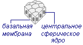
Epithelial tissue(integumentary) - adjacent tissue that covers the body from the outside; lines the internal organs and cavities; is a part of the liver, glands, lungs. In addition, they line the inner surface of blood vessels, airways, and ureters. Epithelial tissues include glandular tissue, which produces various types of secretions (sweat, saliva, gastric juice, pancreatic juice). The cells of this tissue are arranged in the form of a layer, and their peculiarity is their polarity (the upper and lower parts of the cell). Epithelial cells have the ability to restore ( regeneration). There are no blood vessels in the epithelial tissue (cells feed diffusely through the basal lamina).
| Type of fabric (drawing) | Tissue structure | Location | Functions |
| Squamous epithelium
|
| skin surface, oral cavity, esophagus, alveoli, capsules of nephrons, pleura, peritoneum | integumentary, protective, excretory(gas exchange, urine excretion) |
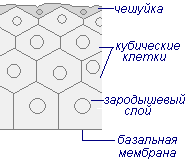
| Cubic epithelium
|
| kidney tubules, salivary glands, endocrine glands | reabsorption (reverse) during the formation of secondary urine, salivation, secretions with hormones |
| Cylindrical epithelium (prismatic)
|
| stomach, intestines, gallbladder, trachea, uterus | mucous membrane of the stomach and intestines |
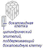
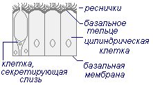
| Unilamellar ciliated epithelium
|
| airways, spinal canal, cerebral ventricles, oviducts | protective(cilia retain and remove dust particles), organizes the flow of fluid, movement of the egg |
| Pseudo-layered
|
| olfactory zones, taste buds of the tongue, urinary canal, trachea | sensitive epithelium... Perception of smell, taste, filling of the bladder, sensation of the presence of foreign particles in the trachea |
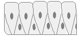
| Multilayer
|
| skin, hair, nails | protective, thermoregulatory, integumentary |
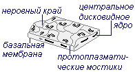
Thus, the following functions are inherent in epithelial tissue: integumentary, protective, trophic, secretory.
Connective tissue
Connective tissue or tissues of the internal environment are represented by blood, lymph and connective tissue. A feature of this tissue is the presence, in addition to cellular elements, of a large amount of intercellular substance, represented by basic substance and fibrous structures(formed by fibrillar proteins - collagen, elastin, etc.). Connective tissue is subdivided into: actually connective, cartilaginous, bone.
Connective tissue proper creates layers of internal organs, subcutaneous tissue, ligaments, tendons and more. Cartilage tissue forms:
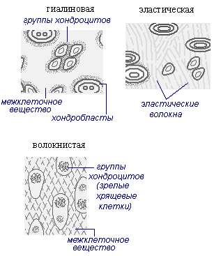
- hyaline cartilage - forms articular surfaces;
- fibrous - located in the intervertebral discs;
- elastic is part of the auricles and epiglottis.
Bone forms the bones of the skeleton, the strength of which is given by deposits of insoluble calcium salts in it. Bone tissue takes part in the mineral metabolism of the body. (See in the section "Musculoskeletal system").
| Type of fabric (drawing) | Tissue structure | Location | Functions |
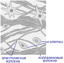
| Loose connective tissue
|
| subcutaneous fatty tissue, pericardial sac, pathways of the nervous system, blood vessels, mesentery | connects skin to muscles, supports organs in the body, fills the gaps between organs. Participates in thermoregulation of the body |
| Cartilage tissue
|
| intervertebral discs, cartilage of the larynx, trachea, ribs, auricle, joint surface, tendon bases, fetal skeleton | smoothing of rubbing surfaces of bones. Protection against deformation of the respiratory tract, ears. Attaching tendons to bones |
Connective tissue functions: protective, supporting, nutritious (trophic).
Muscle cells have the following properties: excitability, contractility, conduction.
Types of muscle tissue
There are three types of muscle tissue: smooth, striated, cardiac.
| Type of fabric (drawing) | Tissue structure | Location | Functions |
| Smooth fabric
|
| forms the muscles of internal organs, is part of the walls of blood and lymphatic vessels | are innervated by the autonomic nervous system and carry out relatively slow movements and tonic contractions |
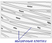
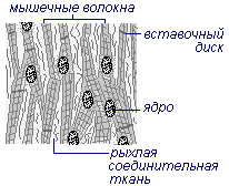
| Striated tissue (muscle fiber)
|
| skeletal muscles, muscles of the tongue, pharynx, the initial part of the esophagus | decrease in response to impulses from the motor neurons of the spinal cord and brain |
| Heart tissue
|
| combines the properties of smooth and striated muscle tissue; heart | responsible for the contraction of all muscle elements |
Functions of muscle tissue: movement of the body in space; displacement and fixation of body parts; changes in the volume of the body cavity, vessel lumen, skin movement; work of the heart.
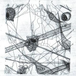
Nerve tissue
Nerve tissue forms the brain and spinal cord, nerve ganglia and fibers. The cells of the nervous tissue are neurons and glial cells. The main feature of neurons is high excitability. They receive irritation (signals) from the external and internal environment of the body, conduct and process them. Neurons are assembled in very complex and numerous circuits that are necessary for receiving, processing, storing and using information.
Types of neurons:
- Unipolar ( motor, centrifugal)
- Pseudo-bipolar ( sensitive, centripetal)
- Multipolar ( part of the brain)
- Dendrites
- Neuron body
- Cell nucleus
- Cytoplasm
- Axons
- Schwann cell
- Axon endings
- Dendron
A neuron consists of cell body(catfish) and two types of processes - dendrites, axons and endplates... In the body of a neuron there is a nucleus with rounded nucleoli.
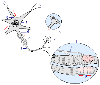
Neuron structure (nerve cell)
- Neuron body
- Dendrites
- Axons
- End plates
- Synaptic vesicles
- Myelin sheath
- Interception of Ranvier
- Nissl's substance
- The end of the nerve fiber
- A section of a muscle fiber that is in a state of contraction
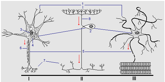
Dendrites(2) - short, thick, highly branching processes that conduct nerve impulses (excitation) to the body of the nerve cell.
Axon(3) - one long (up to 1.5 m) non-branching outgrowth of a nerve cell that conducts a nerve impulse from the cell body to its terminal section. The processes are hollow tubes filled with cytoplasm that flows towards the end plates. The cytoplasm takes up enzymes that were formed in the structures of the granular endoplasmic reticulum (8) and catalyze the synthesis mediators in the end plates (4). Mediators are stored in synaptic vesicles (5). The axons of some neurons are protected from the surface by the myelin sheath (6), formed Schwann cells wrapping around an axon. This shell consists of cells of a kind of nervous tissue - glia, in which all nerve cells are immersed. Glia plays an auxiliary role - it performs an insulating, supporting, trophic and protective function. Locations where the axon is not covered (by the myelin sheath) are called Ranvier interceptions (7). Myelin (a fatty white matter) is a remnant of the membranes of dead cells and its composition provides the insulating properties of the cell.
Nerve cells connect to each other through synapses. Synapse- the place of contact of two neurons, where a nerve impulse is transmitted from one cell to another. Synapses are formed at the points of contact of the axon with the cells to which it transmits information. These areas are somewhat thickened (10), as they contain bubbles of irritating liquid. If nerve impulses reach the synapse, the bubbles burst, the liquid pours out into the synoptic cleft and acts on the membrane of the cell that receives the information. Depending on the composition and amount of biologically active substances contained in the liquid, the cell receiving the information can be excited and intensify its work, or slow down - weaken or stop it altogether.
Cells that receive information usually have many synapses. Through some of them they receive stimulating signals, through others - negative, inhibitory ones. All these signals are summed up, followed by a change in work.
Thus, the functions of the nervous tissue include: receiving, processing, storing, transmitting information coming from the external environment and internal organs; regulation and coordination of the activity of all body systems.
Physiological organ systems
The tissues of the human and animal body form organs and physiological systems of organs: integumentary, support and movement system, digestive, circulatory, respiratory, excretory, sexual, endocrine, nervous.
| Physiological systems | Organs forming the system | Meaning |
| Integumentary system | Skin and mucous membranes | Protects the body from external influences |
| Support and movement system | Bones that make up the skeleton and muscles | Shapes the body, provides support and movement, and protects internal organs |
| Digestive system | Organs of the oral cavity ( tongue, teeth, salivary glands), pharynx, esophagus, stomach, intestines, liver, pancreas | Provides the processing of nutrients in the body |
| Circulatory system | Heart and blood vessels | Carries out the process of blood circulation and metabolism between the body and the environment |
| Respiratory system | Nasal cavity, nasopharynx, trachea, lungs | Provide gas exchange |
| Excretory system | Kidneys, ureters, bladder, urethra | Eliminates the final toxic metabolic products from the body |
| Reproductive system | Male organs(testes, scrotum, prostate, penis). Female organs(ovaries, uterus, vagina, external female genital organs) | Provide reproduction |
| Endocrine system | Endocrine glands (thyroid, genital, pancreas, adrenal glands, etc.) | Produce hormones that regulate functions and metabolism in organs and tissues |
| Nervous system | Nerve tissue that permeates all organs and tissues | Regulates the coordinated functioning of all systems and the whole organism in changing environmental conditions |
Reflex regulation
The nervous system regulates all processes in the body, and also ensures the appropriate response of the body to the effects of the external environment. These functions of the nervous system are performed reflexively. Reflex- the body's response to irritation, which occurs with the participation of the central nervous system. Reflexes are carried out due to the propagation of the excitation process along the reflex arc. Reflex activity is the result of the interaction of two processes - excitation and inhibition.
Excitation and inhibition are two opposite processes, the interaction of which ensures the coordinated activity of the nervous system and the coordinated work of the organs of our body.
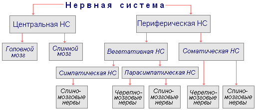
Central and peripheral nervous system
Most neurons are found in the brain and spinal cord. They make up central nervous system(CNS). Some of these neurons go beyond its limits: their long processes are combined into bundles, which, as part of the nerves, go to all organs of the body. The nervous system consists of nerve cells - neurons (there are 25 billion neurons in the brain and 25 million in the periphery.
The central nervous system includes the brain and spinal cord. In addition to nerves, in the brain and not in the central nervous system, there are accumulations of bodies of neurons - these are nerve nodes. Peripheral part of the nervous system includes nerves extending from the brain and spinal cord and nerve nodes located outside the brain and spinal cord. By function, the nervous system is divided into somatic and autonomic nervous systems. Somatic - communicates the body with the external environment (perception of irritations, regulation of movements of the striated muscles, etc.), and vegetative - regulates the metabolism and work of internal organs (heartbeat, vascular tone, peristaltic contractions of the intestines, secretion of various glands, etc. .). both of these systems work in close interaction, but the autonomic nervous system has some independence (autonomy), controlling involuntary functions.
Reflex and reflex arc
The activity of the nervous system is of a reflex nature. Reflex is a natural response of the body to changes in the external or internal environment, carried out by the central nervous system in response to stimulation of receptors. Receptors are nerve endings that receive information about changes in the external and internal environment. Any annoyance ( mechanical, light, sound, chemical, electrical, temperature), perceived by the receptor, is converted into an excitation process. Excitation is transmitted along sensitive - centripetal nerve fibers to the central nervous system, where an urgent process of impulse processing takes place. From here, impulses are directed along the fibers of centrifugal neurons to the executive organs that respond to stimulation.
The reflex arc is the path along which nerve impulses pass from the receptors to the executive organ. For the implementation of any reflex, the coordinated work of all links of the reflex arc is necessary.
Reflex arc diagram.

- External stimulus
- Sensory nerve endings in the skin
- Sensory neuron
- Synapse
- Intercalary neuron
- Synapse ( neuron-to-neuron transmission)
- Motor neuron
In the implementation of any reflex action, excitation processes are involved, causing a certain activity, and the inhibition process, which turns off those nerve centers that interfere with the implementation of reflex actions. The process of inhibition is the opposite of arousal. The interaction of the processes of excitation and inhibition underlies nervous activity, regulation and coordination of functions in the body.
Thus, these both processes ( excitation and inhibition) are closely related to each other, which ensures the coordinated activity of all organs and the whole organism.