The history of the discovery of the cell
Cytology ("cytos" - cell, cell) is the science of the cell. Modern cytology studies: the structure of cells, their formation as elementary living systems, explores the formation of individual cellular components, the processes of cell reproduction, repair, adaptation to environmental conditions, and other processes. In other words, modern cytology is cell physiology.
The development of the theory of the cell is closely connected with the invention of the microscope (from the Greek "micros" - small, "scopeo" - I examine). This is due to the fact that the human eye is unable to distinguish between objects smaller than 0.1 mm, which is 100 micrometers (abbreviated microns or microns). The sizes of cells (and even more so, intracellular structures) are significantly smaller.
For example, the diameter of an animal cell usually does not exceed 20 microns, that of a plant cell is 50 microns, and the length of the chloroplast of a flowering plant is no more than 10 microns. With the help of a light microscope, objects with a diameter of tenths of a micron can be distinguished.
The first microscope was designed in 1610 by Galileo and was a combination of lenses in a lead tube (Fig. 1.1). And before this discovery in 1590, the Dutch craftsmen Jansen were engaged in the manufacture of glasses.
Rice. 1.1. Galileo Galilei (1564-1642)
For the first time, the microscope for research was used by the English physicist and naturalist R. Hooke (Fig. 1.2, 1.4). In 1665, he first described the cellular structure of cork and introduced the term "cell" (Fig. 1.3). R. Hooke made the first attempt to count the number of cells in a certain volume of cork.
He formulated the idea of a cell as a cell completely closed on all sides and established the fact of the cellular structure of plant tissues. These two main conclusions determined the direction of further research in this area.
Rice. 1.2. Robert Hooke (1635-1703)
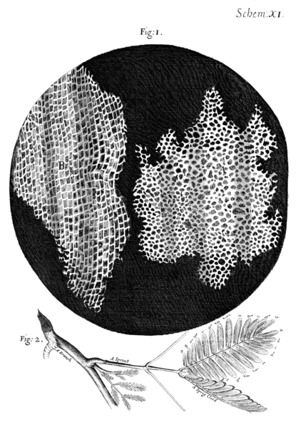
Rice. 1.3. Cork cells studied by Robert Hooke
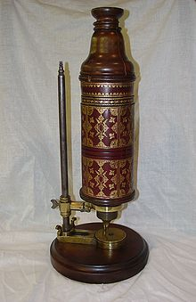
Rice. 1.4. Microscope by Robert Hooke
In 1674, the Dutch merchant Antonio van Leeuwenhoek, using a microscope, first saw “animals” in a drop of water - moving living organisms (single-celled organisms, blood cells, spermatozoa) and reported this to the scientific community (Fig. 1.5, 1.6). The descriptions of these "animalcuses" earned the Dutchman worldwide fame and aroused interest in the study of the living microcosm.
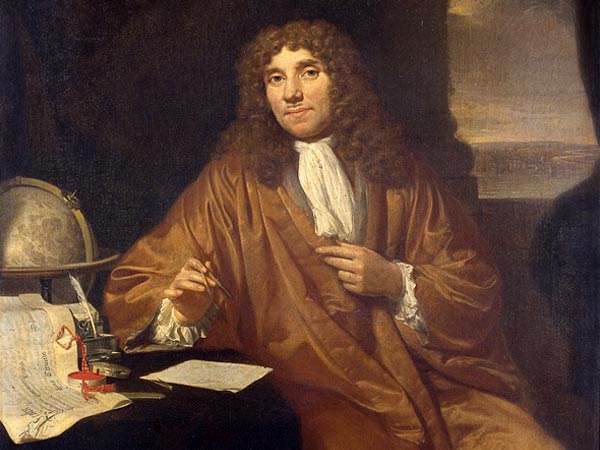
Rice. 1.5. Antonio van Leeuwenhoek (1632-1723)
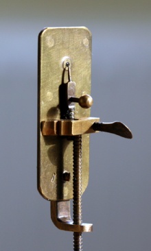
Rice. 1.6. Microscope by Antonio van Leeuwenhoek
In 1693, during the stay of Peter I in Delphi, A. Leeuwenhoek demonstrated to him how blood moves in the fin of a fish. These demonstrations made such a great impression on Peter I that when he returned to Russia, he created a workshop for optical instruments. Petersburg Academy of Sciences was organized in 1725.
Talented masters I.E. Belyaev, I.P. Kulibin made microscopes (Fig. 1.7, 1.8, 1.9), in the design of which academicians L. Euler, F. Epinus took part.
Rice. 1.7. I.P. Kulibin (1735-1818)
Rice. 1.8. I.E. Belyaev
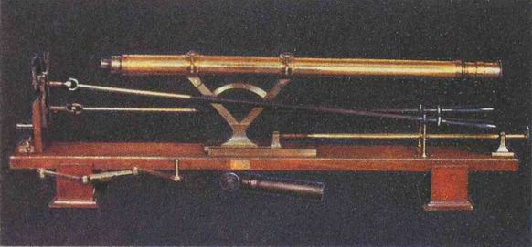
Rice. 1.9. Microscopes made by Russian craftsmen
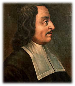
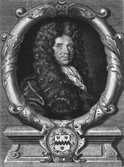
In 1671–1679 Italian biologist and physician Marcello Malpighi gave the first systematic description of the microstructure of plant organs, which marked the beginning of plant anatomy (Fig. 1.10).
Rice. 1.10. Marcello Malpighi (1628-1694)
In 1671–1682 the Englishman Nehemiah Grew described in detail the microstructures of plants; introduced the term "tissue" to refer to the concept of a collection of "vesicles", or "sacs" (Fig. 1.11). Both of these researchers (they worked independently of each other) gave amazingly accurate descriptions and drawings. They came to the same conclusion regarding the generality of the construction of plant tissue from vesicles.
Rice. 1.11. Nehemiah Grew (1641-1712)
In the 20s of the XIX century. the most significant works in the field of studying plant and animal tissues belong to the French scientists Henri Dutrochet (1824), Francois Raspail (1827), Pierre Turpin (1829). They argued that cells (sacs, vesicles) are the elementary structures of all plant and animal tissues. These studies paved the way for the discovery of the cell theory.
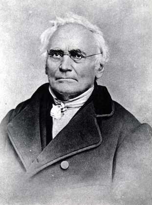
One of the founders of embryology and comparative anatomy, academician of the St. Petersburg Academy of Sciences, Karl Maksimovich Baer, showed that the cell is a unit not only of the structure, but also of the development of organisms (Fig. 1.12).
Rice. 1.12. K.M. Baer (1792-1876)
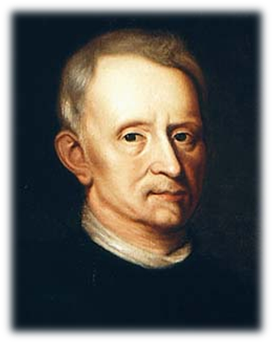
In 1759, the German anatomist and physiologist Caspar Friedrich Wolf proved that the cell is a unit of growth. (Fig. 1.13).
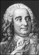
Rice. 1.13. K.F. Wolf (1733–1794)
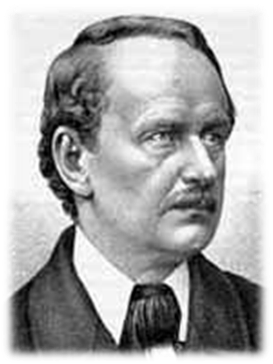
1830s Czech physiologist and anatomist Ya.E. Purkyne (Fig. 1.14), German biologist I.P. Muller proved that cellular organization is universal for all types of tissues.
Rice. 1.14. Ya.E. Purkyne (1787-1869)
In 1833 the British botanist R. Brown (Fig. 1.15) described the nucleus of a plant cell.
Rice. 1.15. Robert Brown (1773-1858)
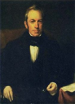
In 1837 Matthias Jakob Schleiden (Fig. 1.16) proposed a new theory of the formation of plant cells, recognizing the decisive role in this process of the cell nucleus. In 1842, he first discovered nucleoli in the nucleus.
According to modern concepts, Schleiden's specific studies contained a number of errors: in particular, Schleiden believed that cells can originate from a structureless substance, and a plant embryo can develop from a pollen tube (the hypothesis of spontaneous generation of life).
Rice. 1.16. Matthias Jacob Schleiden (1804-1881)
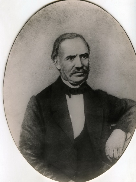
German cytologist, histologist and physiologist Theodor Schwann (Fig. 1.17) got acquainted with the works of the German botanist M. Schleiden, who described the role of the nucleus in the plant cell. Comparing these works with his own observations, Schwann developed his own principles of the cellular structure and development of living organisms.
In 1838, Schwann published three preliminary messages on the cellular theory, and in 1839, the work "Microscopic studies on the correspondence in the structure and growth of animals and plants", where he published the basic principles of the theory of the cellular structure of living organisms.
F. Engels argued that the creation of the cellular theory was one of the three greatest discoveries in the natural sciences of the 19th century, along with the law of energy transformation and evolutionary theory.
Rice. 1.17. Theodor Schwann (1810-1882)
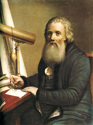
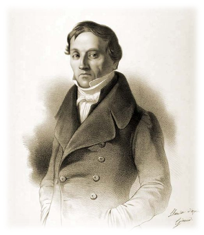
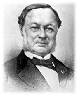
In 1834–1847 Professor of the Medical and Surgical Academy in St. Petersburg P.F. Goryaninov (Fig. 1.18) formulated the principle that the cell is a universal model for the organization of living beings.
Goryaninov divided the world of living beings into two kingdoms: the formless, or molecular, and organic, or cellular. He wrote that "... the organic world is primarily a cellular kingdom ...". He noted in his studies that all animals and plants consist of interconnected cells, which he called bubbles, that is, he expressed an opinion about the general plan of the structure of plants and animals.
Rice. 1.18. P.F. Goryaninov (1796-1865)
In the history of the development of cell theory, two stages can be distinguished:
1) the period of accumulation of observations on the structure of various unicellular and multicellular organisms of plants and animals (about 300 years);
2) the period of generalization of the available data in 1838 and the formulation of the postulates of the cell theory;