The cerebellum has cerebellum, cerebellum
The functions of the cerebellum are similar in various species, including humans. This is confirmed by their disturbance in case of damage to the cerebellum in the experiment in animals and the results of clinical observations in diseases affecting the cerebellum in humans. The cerebellum is a brain center that is extremely important for coordinating and regulating motor activity and maintaining posture. The cerebellum works mainly reflexively, maintaining the balance of the body and its orientation in space. It also plays an important role (especially in mammals) in vlocomotion (moving in space).
Accordingly, the main functions of the cerebellum are:
movement coordination
balance regulation
regulation of muscle tone
ensuring smoothness, rhythm - tactics of movements.
diencephalon
The diencephalon is a part of the brain.
In embryogenesis, the diencephalon is formed on the back of the first cerebral vesicle. In front and above, the diencephalon borders on the anterior, and below and behind - on the midbrain.
The structures of the diencephalon surround the third ventricle.
The diencephalon is subdivided into:
Thalamic brain (Thalamencephalon)
Subthalamic region or hypothalamus (hypothalamus)
The third ventricle, which is the cavity of the diencephalon
Functions of the diencephalon
Movement, including facial expressions.
Metabolism, body temperature, food intake, sleep and wakefulness.
Behavior in extreme situations, manifestations of rage, aggression, pain and pleasure.
Responsible for the feeling of thirst, hunger, satiety.
Instinctive forms of behavior (food, sexual, play, etc.).
All types of sensitivity, except for smell, including sensations of pain, temperature, light touch and pressure, and is also involved in emotional processes and memory.
Short-term and long-term modal non-specific memory.
limbic system is the link between the cerebral cortex and the body. Unity with the body causes physical signs of emotions (blush of shame, smile of joy). The limbic system produces emotions, which in turn either strengthen or weaken the immune system. They directly affect the quality of education, so it is extremely important to reinforce the cognitive processes of children with positive emotions.
The limbic system is made up of five major structures: the thalamus, hypothalamus, amygdala, hippocampus, and basal ganglion.
thalamus works as a "distribution station" for all sensations entering the brain, except for olfactory ones. It also transmits motor impulses from the cerebral cortex through the spinal cord to the musculature. In addition, the thalamus recognizes sensations of pain, temperature, light touch and pressure, and is also involved in emotional processes and memory.
Hypothalamus controls the functioning of the pituitary gland, normal body temperature, food intake, sleep and wakefulness. It is also the center responsible for behavior in extreme situations, manifestations of rage, aggression, pain and pleasure.
amygdala associated with areas of the brain responsible for processing cognitive and sensory information, as well as with areas related to combinations of emotions. The amygdala coordinates reactions of fear or anxiety caused by internal signals.
hippocampus uses sensory information from the thalamus and emotional information from the hypothalamus to form short-term memory. Short-term memory, by activating the nerve networks of the hippocampus, can then move into "long-term storage" and become long-term memory for the entire brain.
Basal ganglion controls nerve impulses between the cerebellum and the anterior lobe of the brain and thereby helps control body movements. It contributes to the control of fine motor skills of the facial muscles and eyes, reflecting emotional states. The basal ganglion is connected to the anterior lobe of the brain through the substantia nigra. It coordinates the thought processes involved in planning the order and coherence of upcoming actions in time.
The processing of all emotional and cognitive information in the limbic system is of a biochemical nature: there is a release of certain neurotransmitters (from lat. transmitto - I transfer; biological substances that cause the conduction of nerve impulses). If cognitive processes proceed against the background of positive emotions, then neurotransmitters such as gamma-aminobutyric acid, acetylcholine, interferon and interglukins are produced. They activate thinking and make memorization more efficient. If the learning processes are built on negative emotions, then adrenaline and cortisol are released, which reduce the ability to learn and memorize.
Development limbic system allows the child to establish social connections. Between the ages of 15 months and 4 years, primitive emotions are generated in the hypothalamus and amygdala: rage, fear, aggression. As the neural networks develop, connections are formed with the cortical (cortical) parts of the temporal lobes responsible for thinking, more complex emotions appear with a social component: anger, sadness, joy, grief. With the further development of nerve networks, connections with the anterior parts of the brain are formed and such subtle feelings as love, altruism, empathy, and happiness develop.
With further development limbic system neural networks connect sensory (visual, auditory, olfactory, gustatory, kinesthetic) and motor circuits with emotions and form memory. It is constructed from neural pathways that link into neural circuits. These schemes are constantly modified and supplemented in an infinite number of combinations. They can be modified, reorganized or reduced for greater efficiency. The circuits are connected to the brain centers where specialized sensory information is processed. For example, the occipital region of the brain is responsible for visual information, while the temporal region is responsible for auditory information. It must be remembered that 90% of the basic circuits are formed in the first five years of a child's life, as is the basic pattern of neural networks., which can then be completed. It is this template that is the material basis of the individuality of thinking, memory, abilities, behavior. The schemes of each person are specific, unique and do not repeat one another.
As the limbic system forms, prerequisites are created for the development imaginations. Albert Einstein believed that "imagination is more important than knowledge, since knowledge speaks about everything that is, and imagination - about everything that will be." Imagination develops on the basis of the synthesis of motor-sensory schemes, emotions and memory (K. Hannaford).
HUMAN BRAIN CORTEX - NEOCORTEX
If you straighten the folds of the neocortex, it will occupy an area of 2500 cm 2. Every 60 seconds, he uses more than 0.5 liters of blood and burns 400 kcal daily. The neocortex makes up only 25% of the total volume of the brain, but contains approximately 85% of all neurons. The mass of the brain is only 2% of the total body weight of a person, but uses 20% of the total blood flow for its own blood supply.
The neocortex consists of gray matter, unmyelinated cell bodies of neurons (myelination is the process of formation of a myelin sheath that covers the high-speed pathways of the central nervous system. Myelin sheaths increase the accuracy and speed of impulse transmission in the nervous system).
The bodies of neurons have unlimited possibilities for the formation of new dendrites (a branching process that receives signals from other neurons, receptor cells, or directly from external stimuli; conducts nerve impulses to the body of a neuron) and reorganization of dendritic networks under the influence of new experience acquired throughout life. It has been established that the neural networks in the adult neocortex contain more than a quadrillion (million billion) connections and can process up to 1000 bits of new information per second. This means that the number of signals that can simultaneously be transmitted through the synapses (connections) of the brain exceeds the number of atoms in the known region of the universe.
The doctrine of the structural features of the structure of the cortex is called architectonics.
Cells of the cortex of large hemispheres are less specialized than neurons in other parts of the brain; nevertheless, certain groups of them are anatomically and physiologically closely related to certain specialized parts of the brain. The microscopic structure of the cerebral cortex is not the same in its different parts. These morphological differences in the cortex made it possible to distinguish individual cortical cytoarchitectonic fields. There are several options for classifying cortical fields. Most researchers distinguish 50 cytoarchitectonic fields (for example, according to Brodman).
DO NOT MIX THE CONCEPT OF CYTOARCHITECTONIC FIELDS WITH THE FIELDS OF THE BRAIN CORTEX (PRIMARY, SECONDARY AND TERTIARY FIELDS).
The microscopic structure of the cortex is quite complex. The cortex consists of a number of layers of cells and their fibers.
The main type of crust structure is six-layered, but it is not uniform everywhere. There are areas of the cortex where one of the layers is very pronounced, and the other is weak. In other areas of the crust, some layers are subdivided into sublayers, and so on.
It has been established that the areas of the cortex associated with a certain function have a similar structure. Areas of the cortex, which are close in animals and humans in terms of their functional significance, have a certain similarity in structure. Those areas of the brain that perform purely human functions (speech) are present only in the human cortex, while animals, even monkeys, are absent.
Morphological and functional heterogeneity of the cerebral cortex allowed highlight the centers of vision, hearing, touch, etc., which have their own specific localization. However, it is wrong to sayabout the cortical center as a strictly limited group of neurons. It must be remembered that the specialization of cortical areas is formed in the process of life. In early childhood, the functional areas of the cortex overlap each other, so their boundaries are vague and indistinct. Only in the process of learning, accumulation of own experience in practical activities there is a gradual concentration of functional zones in centers separated from each other.
HORIZONTAL AND VERTICAL CONNECTIONS OF THE BRAIN
White matter of the cerebral hemispheres consists of nerve conductors. In accordance with the anatomical and functional features of the white matter fibers are divided into associative, commissural and projection. Associative fibers unite different parts of the cortex within one hemisphere. These fibers are short and long. Short fibers are usually arcuate and connect adjacent gyri. Long fibers connect distant parts of the cortex.
It is customary to call commissural fibers those fibers that connect topographically identical parts of the right and left hemispheres. Commissural fibers form three commissures: the anterior white commissure, the commissure of the fornix, and the corpus callosum. The anterior white commissure connects the olfactory regions of the right and left hemispheres. The fornix commissure connects the hippocampal gyri of the right and left hemispheres. The bulk of the commissural fibers pass through corpus callosum, connecting symmetrical parts of both hemispheres of the brain.
It is customary to call projection fibers those fibers that connect the hemispheres of the brain with the underlying parts of the brain - the trunk and spinal cord. As part of the projection fibers, there are conducting paths that carry afferent (sensitive) and efferent (motor) information.
Pathways of the brain
In the white matter of the trunk brain and spinal cord the conductors of the ascending and descending directions are located. Descending paths conduct motor impulses from the cerebral cortex (pyramidal path) to the reflex apparatus of the spinal cord, as well as impulses that contribute to the implementation of a motor act (extrapyramidal paths) from various parts of the subcortical formations and the brain stem.
Descending motor conductors end on the peripheral motor neurons of the spinal cord in segments. The overlying sections of the central nervous system have a significant impact on the reflex activity of the spinal cord. They inhibit the reflex mechanisms of the spinal cord's own apparatus. So, with a pathological shutdown of the pyramidal pathways, the own reflex mechanisms of the spinal cord are disinhibited. This increases the reflexes of the spinal cord and muscle tone.
In addition, protective reflexes are detected and those that are normally observed only in newborns and children in the first months of life.
The ascending pathways transmit sensitive impulses from the periphery (from the skin, mucous membranes, muscles, joints, etc.) from the spinal cord to the overlying parts of the brain. Eventually these impulses reach the cerebral cortex. From the periphery, impulses come to the cerebral cortex in two ways: through the so-called specific systems of conductors (through the ascending conductor and thalamus) and in a non-specific system - through the reticular formation (network formation) of the brain stem. All sensitive conductors give off collaterals of the reticular formation. The reticular formation activates cerebral cortex, spreading impulses to different parts of the cortex. Its influence on the cortex is diffuse, while specific conductors send impulses only to certain projection zones.
In addition, the reticular formation is involved in the regulation of various vegetative-visceral and sensorimotor functions of the body. Thus, the overlying parts of the brain are under the influence of the spinal cord.
Mental processes are carried out by complex systems - jointly working zones of the cortex and underlying nervous structures. These lower structures are involved in the work of the cortex, regulating and providing its tone. The data obtained in modern anatomical and physiological studies allow us to formulate the principle of the vertical structure of the functional systems of the brain : each form of behavior is provided by different levels of the nervous system, connected to each other both horizontal (transcortical - commissural and associative) connections, and vertical (top-down and bottom-up - projection). All this turns the brain into a self-regulating system..
association fibers; commissural fibers; projection fibers
The cerebellum is located in the posterior cranial fossa above the medulla oblongata and the pons. Above the cerebellum are the occipital lobes of the cerebrum (see); between them and the cerebellum, the cerebellum is stretched (or baited) - a process of the dura mater.
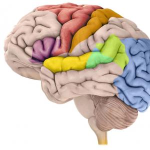
Anatomy and Physiology. In the cerebellum, the upper and lower surfaces, the anterior and posterior edges are distinguished. The cerebellum consists of a middle section, or worm, and two hemispheres, each of which is divided by grooves into three lobes (Fig.). Each lobe of the hemisphere corresponds to a certain part of the worm.
Cerebellum (structure): a - upper, or posterior, surface; b - lower, or front, surface; c - sagittal section through the worm. 1 - front notch; 2 - right hemisphere; 3 - back notch; 4 - left hemisphere; 5 - middle legs of the cerebellum: 6 - shred; 7 - tonsil; 8 - cerebellar worm; 9 - plate of the quadrigemina; 10 - front cerebral sail; 11 - white matter; 12 - cerebellar cortex; 13 - medulla oblongata; 14 - bridge.
In the cerebellum, there is a white matter embedded inside and a gray cortical substance covering it with a thin layer. The white matter of the cerebellar hemispheres connects medially to the white matter of the vermis. The picture of the location of the white matter, visible on the sagittal section of the worm, due to its similarity with the picture of the branching of the tree, is called the tree of life. In the white matter there are also accumulations of gray matter - the nuclei of the cerebellum, of which the more important are the jagged nuclei of the roof and the nuclei of the tent.
The white matter of the cerebellar hemispheres is connected to neighboring parts of the brain through fibrous bundles. These bundles form strands of varying thickness, called cerebellar peduncles, and connect the cerebellum to the pons, to the midbrain and medulla oblongata.
The middle legs exit the cerebellum laterally and, gradually approaching, go forward, passing into the bridge.
The upper, or front, legs are located medially from the middle ones, go forward and disappear in the form of flattened round strands (also gradually converging) under the quadrigemina, in the area of the red nuclei. Between them is placed the front cerebral sail.
The lower legs go back and down to the medulla oblongata.
The main function of the cerebellum is the regulation of the coordinated (coordinated) activity of skeletal muscles.
Together with the cerebral cortex, the cerebellum is involved in the coordination of so-called voluntary movements. This is done thanks to the connections of the cerebellum with those embedded in the skeletal muscles, joints and tendons.
Together with the vestibular apparatus of the semicircular canals of the inner ear (see), signaling to the central nervous system about the position of the head and body in space, the cerebellum is involved in the regulation of body balance (see) when walking and active movements.
The cerebellum regulates the coordination of skeletal muscle movements through special systems of conductive fibers running from the cerebellum to the anterior horns, where the peripheral motor nerves of skeletal muscles originate.
Pathology. When the cerebellum is damaged, mainly disorders of the coordinated activity of skeletal muscles develop, namely: impaired coordination of voluntary movements and imbalance of the body. The first group of cerebellar movement disorders is manifested in violations of the smooth movements of the limbs (mainly hands) and, in particular, in the appearance (see) at the end of a purposeful movement; c (the so-called scanned, in which not semantic, but rhythmic arrangement of stresses in words appears); in the slowness of voluntary movements and speech; in changing handwriting. Cerebellar imbalances are manifested mainly in dizziness and a change in gait (see Ataxia), which takes on the character of a drunken gait, and the patient staggers towards the lesion. All of these disorders are sometimes accompanied by nystagmus (twitching of the eyeballs when they are retracted).
A common symptom of damage to the cerebellum is a disorder in the coordinated activity of muscles belonging to different muscle groups, with their participation in one motor act.
Such asynergy of the muscles of the legs and torso is manifested, for example, when a patient tries to take a sitting position from a lying position without the help of hands.
Among tumors of the cerebellum, infiltratively growing benign neoplasms, astrocytomas, and angioreticulomas are most common.

Medulloblastoma
Of the malignant tumors of the cerebellum, the first place belongs to medulloblastomas, sarcomas. Tumors of the cerebellum are subject to surgical treatment. When open and there may be mechanical damage to the cerebellar tissue. compression by a large focal hematoma located in the posterior cranial fossa. In this case, surgical intervention with removal of the hematoma is indicated.
In some cases, after suffering meningitis, after the resorption of a hemorrhage of traumatic origin, atrophy of the cerebellum develops.
Surgical treatment of diseases of the cerebellum. Operations on the cerebellum are performed with its tumors, abscesses, cysts, hemorrhages, traumatic injuries. The patient's position on
7.1. STRUCTURE, CONNECTIONS AND FUNCTIONS OF THE CERENELS
The cerebellum (cerebellum) is located under the duplication of the dura mater, known as outline of the cerebellum(tentorium cerebelli), which divides the cranial cavity into two unequal spaces - supratentorial and subtentorial. V subtentorial space, the bottom of which is the posterior cranial fossa, in addition to the cerebellum, there is a brain stem. The volume of the cerebellum averages 162 cm 3 . Its mass varies within 136-169 g.
The cerebellum is located above the pons and medulla oblongata. Together with the upper and lower medullary sails, it makes up the roof of the fourth ventricle of the brain, the bottom of which is the so-called rhomboid fossa (see Chapter 9). Above the cerebellum are the occipital lobes of the cerebrum, separated from it by the indentation of the cerebellum.
The cerebellum is divided into two hemispheres(hemispherum cerebelli). Between them in the sagittal plane above the fourth ventricle of the brain is the phylogenetically most ancient part of the cerebellum - its worm(vermis cerebelli). The vermis and cerebellar hemispheres are fragmented into lobules by deep transverse grooves.
The cerebellum consists of gray and white matter. The gray matter forms the cerebellar cortex and the paired nuclei of nuclei cerebelli located in its depth (Fig. 7.1). The largest of them - jagged nuclei(nucleus dentatus) - located in the hemispheres. In the central part of the worm there are tent cores(nuclei
Rice. 7.1. Cerebellar nuclei.
1 - dentate nucleus; 2 - cork-like nucleus; 3 - the core of the tent; 4 - spherical nucleus.

Rice. 7.2.Sagittal section of the cerebellum and brainstem.
1 - cerebellum; 2 - "tree of life"; 3 - front cerebral sail; 4 - plate of the quadrigemina; 5 - aqueduct of the brain; 6 - leg of the brain; 7 - bridge; 8 - IV ventricle, its choroid plexus and tent; 9 - medulla oblongata.
fastigii), between them and the dentate nuclei are spherical and corky nuclei(nuctei. globosus et emboliformis).
Due to the fact that the cortex covers the entire surface of the cerebellum and penetrates into the depth of its furrows, on the sagittal section of the cerebellum, its tissue has a leaf pattern, the veins of which are formed by white matter (Fig. 7.2), which constitutes the so-called cerebellum tree of life (arbor vitae cerebelli). At the base of the tree of life is a wedge-shaped notch, which is the upper part of the cavity of the IV ventricle; the edges of this notch form his tent. The cerebellar worm serves as the roof of the tent, and its front and back walls are made up of thin cerebral plates, known as the anterior and posterior brain sails(vella medullare anterior et posterior).
Some information about architectonics of the cerebellum, giving grounds for judging the function of its components. At cerebellar cortex There are two cell layers: the inner one is granular, consisting of small granule cells, and the outer one is molecular. Between them is a number of large pear-shaped cells, bearing the name of the Czech scientist I. Purkinje (Purkinje I., 1787-1869) who described them.
Impulses enter the cerebellar cortex through the mossy and creeping fibers penetrating into it from the white matter, which make up the afferent pathways of the cerebellum. Mossy fibers carry impulses from the spinal cord
vestibular nuclei and pontine nuclei are transmitted to the cells of the granular layer of the cortex. The axons of these cells, together with creeping fibers passing through the granular layer in transit and carrying impulses from the lower olives to the cerebellum, reach the superficial, molecular layer of the cerebellum. Here, the axons of the cells of the granular layer and the creeping fibers divide in a T-shape, and in the molecular layer their branches take a direction longitudinal to the surface of the cerebellum. The impulses that have reached the molecular layer of the cortex, passing through the synaptic contacts, fall on the branching of the dendrites of the Purkinje cells located here. Then they follow the dendrites of Purkinje cells to their bodies located on the border of the molecular and granular layers. Then, along the axons of the same cells crossing the granular layer, they penetrate into the depths of the white matter. The axons of Purkinje cells terminate in the nuclei of the cerebellum. Mainly in the dentate nucleus. Efferent impulses coming from the cerebellum along the axons of the cells that make up its nuclei and take part in the formation of the cerebellar peduncles leave the cerebellum.
The cerebellum has three pairs of legs: bottom, middle and top. The lower leg connects it with the medulla oblongata, the middle one with the bridge, the upper one with the midbrain. The peduncles of the brain make up pathways that carry impulses to and from the cerebellum.
The cerebellar vermis provides stabilization of the center of gravity of the body, its balance, stability, regulation of the tone of reciprocal muscle groups, mainly the neck and trunk, and the emergence of physiological cerebellar synergies that stabilize the balance of the body.
To successfully maintain the balance of the body, the cerebellum constantly receives information passing through the spinocerebellar pathways from the proprioceptors of various parts of the body, as well as from the vestibular nuclei, inferior olives, the reticular formation and other formations involved in controlling the position of body parts in space. Most of the afferent pathways leading to the cerebellum pass through the inferior cerebellar peduncle, some of them are located in the superior cerebellar peduncle.
proprioceptive impulses, going to the cerebellum, like other sensitive impulses, following the dendrites of the first sensitive neurons, they reach their bodies located in the spinal nodes. Subsequently, the impulses going to the cerebellum along the axons of the same neurons are directed to the bodies of the second neurons, which are located in the inner sections of the base of the posterior horns, forming the so-called Clark's pillars. Their axons enter the lateral divisions of the lateral cords of the spinal cord, where they form spinal tracts, in this case, part of the axons enters the lateral column of the same side and forms there posterior spinocerebellar tract Flexig (tractus spinocerebellaris posterior). Another part of the axons of the cells of the posterior horns passes to the other side of the spinal cord and enters the opposite lateral funiculus, forming in it anterior spinocerebellar tract of Gowers (tractus spinocerebellaris anterior). The spinal tracts, increasing in volume at the level of each spinal segment, rise to the medulla oblongata.
In the medulla oblongata, the posterior spinocerebellar tract deviates laterally and, having passed through the inferior cerebellar peduncle, penetrates into the cerebellum. The anterior spinocerebellar tract passes through the medulla oblongata, the pons of the brain and reaches the midbrain, at the level of which it makes its second decussation in the anterior medullary velum and passes into the cerebellum through the superior cerebellar peduncle.
Thus, of the two spinal tracts, one never crosses (non-crossed Flexig's path), and the other goes to the opposite side twice (doubly crossed Gowers' path). As a result, both conduct impulses from each half of the body, mainly to the homolateral half of the cerebellum.
In addition to the spinal cerebellar pathways of Flexig, impulses to the cerebellum pass through the inferior cerebellar peduncle through vestibulocerebellar tract (tractus vestibulocerebellaris), which begins mainly in the upper vestibular nucleus of Bechterew, and along olivocerebellar tract (tractus olivocerebellaris), coming from the lower olive. Part of the axons of the cells of the thin and wedge-shaped nuclei, not taking part in the formation of the bulbothalamic tract, in the form of external arcuate fibers (fibre arcuatae externae) also enters the cerebellum through the inferior cerebellar peduncle.
Through its middle legs, the cerebellum receives impulses from the cerebral cortex. These impulses travel through cortical-cerebellopontine pathways, consisting of two neurons. The bodies of the first neurons are located in the cerebral cortex, mainly in the cortex of the posterior sections of the frontal lobes. Their axons pass as part of the radiant crown, the anterior leg of the internal capsule and end in the nuclei of the bridge. Axons of cells of the second neurons, whose bodies are located in their own nuclei of the bridge, go to its opposite side and make up the middle cerebellar peduncle after decussation,
ending in the opposite hemisphere of the cerebellum.
Part of the impulses that have arisen in the cerebral cortex of the brain reaches the opposite hemisphere of the cerebellum, bringing information not about the produced, but only about the active movement planned for execution. Having received such information, the cerebellum instantly sends out impulses that correct voluntary movements, mainly, by repaying inertia and the most rational regulation of the tone of reciprocal muscles - agonist and antagonist muscles. As a result, a kind of eimetry, making arbitrary movements clear, polished, devoid of inappropriate components.
The pathways leaving the cerebellum consist of axons of cells whose bodies form its nuclei. Most efferent pathways, including those from the dentate nuclei, leave the cerebellum through its superior peduncle. At the level of the inferior tubercles of the quadrigemina, the efferent cerebellar tracts are crossed (Crossing of the superior cerebellar peduncles of Wernecking). After the cross, each of them reaches the red nuclei of the opposite side of the midbrain. In the red nuclei, the cerebellar impulses switch to the next neuron and then move along the axons of cells whose bodies are embedded in the red nuclei. These axons form in red nuclear-spinal pathways (tracti rubro spinalis), Monakov's ways, which shortly after exits from the red nuclei are subjected to crossover (tire cross or Trout cross), after which they descend into the spinal cord. In the spinal cord, the red nuclear spinal cords are located in the lateral cords; their constituent fibers terminate at the cells of the anterior horns of the spinal cord.
The entire efferent pathway from the cerebellum to the cells of the anterior horns of the spinal cord can be called cerebellar-red-nuclear-spinal (tractus cerebello-rubrospinalis). He crosses twice (decussation of the superior cerebellar peduncles and decussation of the tegmentum) and ultimately connects each hemisphere of the cerebellum with peripheral motor neurons located in the anterior horns of the homolateral half of the spinal cord.
From the nuclei of the cerebellar vermis, the efferent pathways go mainly through the inferior cerebellar peduncle to the reticular formation of the brainstem and the vestibular nuclei. From here, along the reticulospinal and vestibulospinal tracts, passing through the anterior cords of the spinal cord, they also reach the cells of the anterior horns. Part of the impulses coming from the cerebellum, passing through the vestibular nuclei, enters the medial longitudinal bundle, reaches the nuclei of the III, IV and VI cranial nerves, which provide movement of the eyeballs, and affects their function.
Summing up, it is necessary to emphasize the following:
1. Each half of the cerebellum receives impulses mainly a) from the homolateral half of the body, b) from the opposite hemisphere of the brain, which has cortico-spinal connections with the same half of the body.
2. From each half of the cerebellum, efferent impulses are sent to the cells of the anterior horns of the homolateral half of the spinal cord and to the nuclei of the cranial nerves that provide movement of the eyeballs.
This nature of the cerebellar connections makes it possible to understand why, with damage to one half of the cerebellum, cerebellar disorders occur predominantly in the same, i.e. homolateral, half of the body. This is especially clearly manifested in the defeat of the cerebellar hemispheres.
7.2. STUDY OF THE FUNCTIONS OF THE CEREbellUM
AND CLINICAL MANIFESTATIONS OF ITS DEFEAT
With damage to the cerebellum, disorders of statics and coordination of movements, muscle hypotension and nystagmus are characteristic.
cerebellar injury, primarily his worm leads to violations of statics - the ability to maintain a stable position of the center of gravity of the human body, balance, stability. When this function is disturbed, static ataxia (from the Greek ataxia - disorder, instability). The patient's instability is noted. Therefore, in a standing position, he spreads his legs wide, balances with his hands. Especially clearly static ataxia is detected with an artificial decrease in the area of support, in particular in the Romberg position. The patient is asked to stand up with his feet firmly together and slightly raising his head. In the presence of cerebellar disorders, the patient is unstable in this position, his body sways, sometimes he is “pulled” in a certain direction, and if the patient is not supported, he may fall. In the case of damage to the cerebellar vermis, the patient usually sways from side to side and often falls back. With pathology of the cerebellar hemisphere, there is a tendency to fall mainly towards the pathological focus. If the static disorder is moderately expressed, it is easier to identify it in the so-called complicated or sensitized Romberg position. The patient is asked to put his feet in line so that the toe of one foot rests on the heel of the other. The assessment of stability is the same as in the usual Romberg position.
Normally, when a person is standing, the muscles of his legs are tense. (support reaction), with the threat of falling to the side, his leg on that side moves in the same direction, and the other leg comes off the floor (jump reaction). With damage to the cerebellum (mainly the worm), the patient's reactions are disturbed
support and jump. Violation of the support reaction is manifested by the instability of the patient in a standing position, especially in the Romberg position. Violation of the jump reaction leads to the fact that if the doctor, standing behind the patient and insuring him, pushes the patient in one direction or another, then the patient falls with a small push (symptom of pushing).
With damage to the cerebellum, the patient's gait is usually changed due to the development statolocomotor ataxia. "Cerebellar" gait in many ways resembles the walk of a drunk person, which is why it is sometimes called the "drunk walk". The patient, due to instability, walks unsteadily, legs wide apart, while he is “thrown” from side to side. And when the hemisphere of the cerebellum is damaged, it deviates when walking from a given direction towards the pathological focus. Particularly pronounced instability when cornering. If the ataxia is pronounced, then the patients completely lose the ability to control their body and cannot not only stand and walk, but even sit.
The predominant lesion of the cerebellar hemispheres leads to a breakdown of its counter-inertial influences, in particular to the occurrence of kinetic ataxia. It is manifested by awkwardness of movements and is especially pronounced with movements that require precision. To identify kinetic ataxia, tests are carried out for coordination of movements. The following is a description of some of them.
Diadochokinesis test (from Greek diadochos - sequence). The patient is invited to close his eyes, stretch his arms forward and quickly, rhythmically supinate and pronate the hands. In case of damage to the hemisphere of the cerebellum, the movements of the hand on the side of the pathological process turn out to be more sweeping (a consequence of dysmetria, more precisely, hypermetry), as a result, the hand begins to lag behind. This indicates the presence of adiadochokinesis.
Finger test. The patient with his eyes closed should take his hand away, and then, slowly, with his index finger, touch the tip of the nose. In the case of cerebellar pathology, the hand on the side of the pathological focus makes an excessive movement in terms of volume (hypermetry), as a result, the patient misses. With a finger-to-nose test, a characteristic of cerebellar pathology is revealed cerebellar (intentional) tremor, whose amplitude increases as the finger approaches the target. This test allows you to identify the so-called bradytelkinesia (symptom of a bridle): near the target, the movement of the finger slows down, sometimes even stops, and then resumes again.
Finger-finger test. The patient with his eyes closed is asked to spread his hands wide and then bring the index fingers together, trying to get the finger into the finger, while, as in the finger-nose test, intentional trembling and a symptom of a bridle are detected.
Heel-knee test (Fig. 7.3). The patient, lying on his back with his eyes closed, is offered to raise one leg high and then hit the knee of the other leg with his heel. With cerebellar pathology, the patient cannot or it is difficult for him to get his heel into the knee of the other leg, especially when performing the test with the leg homolateral to the affected hemisphere of the cerebellum. If, nevertheless, the heel reaches the knee, then it is proposed to hold it, slightly touching the front surface of the lower leg, down to the ankle joint, while in the case of cerebellar pathology, the heel always slides off the lower leg in one direction or the other.

Rice. 7.3.Heel-knee test.
Index test: The patient is invited several times with his index finger to hit the rubber tip of the hammer, which is in the examiner's hand. In the case of cerebellar pathology in the patient's hand on the side of the affected cerebellar hemisphere, a miss due to dysmetria is noted.
Symptom of Thomas-Jumenty: If the patient takes an object, such as a glass, he spreads his fingers excessively.
Cerebellar nystagmus. Twitching of the eyeballs when looking to the side (horizontal nystagmus) is considered as a consequence of intentional trembling of the eyeballs (see chapter 30).
Speech disorder: Speech loses its smoothness, becomes explosive, fragmented, chanted like cerebellar dysarthria (see Chapter 25).
Handwriting change: In connection with the disorder of coordination of hand movements, the handwriting becomes uneven, the letters are deformed, excessively large (megalography).
Pronator Phenomenon: The patient is asked to hold the arms outstretched forward in the supination position, while spontaneous pronation soon occurs on the side of the affected cerebellar hemisphere.
Goff-Schilder's symptom: If the patient holds his arms outstretched forward, then on the side of the affected hemisphere, the arm is soon retracted outward.
simulation phenomenon. The patient with his eyes closed should quickly give his hand a position similar to that which the examiner had previously given to his other hand. When the hemisphere of the cerebellum is affected, the arm homolateral to it makes a movement that is excessive in amplitude.
The Doinikov phenomenon. finger phenomenon. The seated patient is asked to place supinated hands with spread fingers on their thighs and close their eyes. In the case of damage to the cerebellum on the side of the pathological focus, spontaneous flexion of the fingers and pronation of the hand and forearm soon occur.
Stuart-Holmes symptom. The examiner asks the patient sitting on a chair to bend the supinated forearms and at the same time, taking his hands by the wrists, resists him. If, at the same time, the patient's hands are suddenly released, then the hand on the side of the lesion, bending by inertia, will hit him in the chest with force.
Muscle hypotension. Damage to the cerebellar vermis usually leads to diffuse muscular hypotension. With damage to the cerebellar hemisphere, passive movements reveal a decrease in muscle tone on the side of the pathological process. Muscle hypotension leads to the possibility of overextension of the forearm and lower leg (symptom of Olshansky) with passive movements, to the appearance dangling hand or foot symptoms when they are passively shaken.
Pathological cerebellar asynergia. Violations of physiological synergies during complex motor acts are detected, in particular, during the following tests (Fig. 7.4).
1. Asynergy according to Babinsky in a standing position. If a patient standing with shifted legs tries to bend back, throwing his head back, then normally in this case there is a flexion of the knee joints. With cerebellar pathology, due to asynergy, this friendly movement is absent, and the patient, losing balance, falls back.

Rice. 7.4.Cerebellar asynergy.
1 - gait of a patient with severe cerebellar ataxia; 2 - back tilt of the body is normal; 3 - with damage to the cerebellum, the patient, leaning back, cannot maintain balance; 4 - performing a test for cerebellar asynergy according to Babinsky by a healthy person; 5 - performing the same test in patients with cerebellar lesions.
2. Asynergy according to Babinsky in the prone position. The patient, lying on a hard plane with outstretched legs, divorced to the width of the shoulder girdle, is invited to cross his arms over his chest and then sit down. In the presence of cerebellar pathology, due to the absence of friendly contraction of the gluteal muscles (manifestation of asynergy), the patient cannot fix the legs and pelvis on the support area, as a result, the legs rise and he fails to sit down. The significance of this symptom in elderly patients, in people with a flabby or obese abdominal wall, should not be overestimated.
Summarizing the foregoing, the diversity and importance of the functions performed by the cerebellum should be emphasized. As part of a complex feedback regulatory mechanism, the cerebellum acts as a focal point for balancing the body and maintaining muscle tone. As P. Duus (1995) notes, the cerebellum provides the ability to perform discrete and precise movements, at the same time, the author reasonably believes that the cerebellum works like a computer, tracking and coordinating sensory information at the input and modeling motor signals at the output.
7.3. MULTISYSTEM DEGENERATIONS
WITH SIGNS OF CEREBELLULAR PATHOLOGY
Multisystemic degenerations are a group of neurodegenerative diseases, the common feature of which is the multifocal nature of the lesion with the involvement of various functional and neurotransmitter systems of the brain in the pathological process and, therefore, the polysystemic clinical manifestations.
7.3.1. Spinal ataxia
Spino-cerebellar ataxias are progressive hereditary degenerative diseases in which the structures of the cerebellum, the brain stem and the pathways of the spinal cord, mainly related to the extrapyramidal system, are mainly affected.
7.3.1.1. Friedreich's hereditary ataxia
Hereditary disease described in 1861 by the German neurologist N. Friedreich (Friedreich N., 1825-1882). It is inherited in an autosomal recessive manner or (less commonly) in an autosomal dominant pattern with incomplete penetrance and variable gene expression. Sporadic cases of the disease are also possible.
Pathogenesisdisease is not specified. In particular, there is no idea of the primary biochemical defect that forms its basis.
Pathomorphology.Pathological anatomical studies revealed a pronounced thinning of the spinal cord, due to atrophic processes in its posterior and lateral cords. As a rule, the sphenoid (Burdach) and tender (Goll) pathways and the spinal cerebellar pathways of Gowers and Flexig, as well as the crossed pyramidal pathway containing
many fibers related to the extrapyramidal system. Degenerative processes are also expressed in the cerebellum, in its white matter and nuclear apparatus.
Clinical manifestations. The disease manifests itself in children or young people under the age of 25 years. S.N. Davidenkov (1880-1961) noted that more often clinical signs of the disease occur in children of 6-10 years of age. The first sign of the disease is usually ataxia. Patients experience uncertainty, staggering when walking, gait changes (when walking, legs are widely spaced). The gait in Friedreich's disease can be called tabetic-cerebellar, since its changes are due to a combination of sensitive and cerebellar ataxia, as well as a usually pronounced decrease in muscle tone. Disorders of statics, discoordination in the hands, intentional tremor, and dysarthria are also characteristic. Possible nystagmus, hearing loss, elements of scant speech, signs of pyramidal insufficiency (tendon hyperreflexia, pathological foot reflexes, sometimes some increase in muscle tone), imperative urge to urinate, decreased sexual potency. Sometimes hyperkinesis of an athetoid nature appears.
An early disorder of deep sensitivity leads to a progressive decrease in tendon reflexes: first on the legs, and then on the hands. Over time, hypotrophy of the muscles of the distal legs is formed. The presence of anomalies in the development of the skeleton is characteristic. First of all, this is manifested by the presence Friedreich's feet the foot is shortened, "hollow", with a very high arch. The main phalanges of her fingers are unbent, the rest are bent (Fig. 7.5). Possible deformation of the spine, chest. Often there are manifestations of cardiopathy. The disease progresses slowly, but steadily leads to disability of patients who eventually become bedridden.
Treatment. Pathogenetic treatment has not been developed. Assign drugs that improve metabolism in the structures of the nervous system, tonic. With severe deformity of the feet, orthopedic shoes are indicated.

Rice. 7.5.Friedreich's foot.
7.3.1.2. Hereditary cerebellar ataxia (Pierre Marie's disease)
This is a chronic progressive hereditary disease that manifests itself at the age of 30-45 years, with slowly increasing cerebellar disorders in combination with signs of pyramidal insufficiency, while characterized by static and dynamic cerebellar ataxia, intentional trembling, chanted speech, tendon hyperreflexia. Possible clonuses, pathological pyramidal reflexes, strabismus, decreased vision, narrowing of the visual fields due to primary atrophy of the optic nerves and pigmentary degeneration of the retina. The course of the disease is slowly progressive. There is a decrease in the size of the cerebellum, cell degeneration
Purkinje, inferior olives, spinal tracts. It is inherited in an autosomal dominant manner. The disease was described in 1893 by the French neurologist R. Marie (1853-1940).
Currently, there is no unanimity in the understanding of the term "Pierre Marie's disease", and the question of the possibility of separating it into an independent nosological form is debatable.
Treatment has not been developed. Usually used metabolically active and tonic, as well as symptomatic agents.
7.3.2. Olivopontocerebellar dystrophy (Dejerine-Thomas disease)
This is a group of chronic progressive hereditary diseases in which dystrophic changes develop mainly in the cerebellum, inferior olives, in the pontine nuclei and in the brain structures associated with them.
With the development of the disease at a young age, about half of the cases are inherited in a dominant or recessive manner, the rest are sporadic. In sporadic cases of the disease, manifestations of akinetic-rigid syndrome and progressive autonomic failure are more common. The average age of the patient with the manifestation of the hereditary form of the disease in the phenotype is 28 years, with the sporadic form of the disease - 49 years, the average life expectancy is 14.9 and 6.3 years, respectively. In the sporadic form, in addition to atrophy of the olives, pons and cerebellum, lesions of the lateral funiculi of the spinal cord, substantia nigra and striatum, a bluish spot in the rhomboid fossa of the fourth ventricle of the brain are more often found.
Symptoms of the growing cerebellar syndrome are characteristic. Sensitivity disorders, elements of bulbar and akinetic-rigid syndromes, hyperkinesis, in particular myorhythmias in the uvula and soft palate, ophthalmoparesis, decreased visual acuity, and intellectual disorders are possible. The disease was described in 1900 by French neuropathologists J. Dejerine and A. Thomas.
The disease often debuts with violations when walking - instability, discoordination, unexpected falls are possible. These disorders may be the only manifestation of the disease for 1-2 years. In the future, coordinating disorders in the hands arise and grow: manipulations with small objects are difficult, handwriting is disturbed, intentional tremor occurs. Speech becomes intermittent, blurred, with a nasal tinge and a breathing rhythm that does not correspond to the construction of speech (the patient speaks as if he was being strangled). At this stage of the disease, manifestations of progressive vegetative insufficiency join, signs of an akinetic-rigid syndrome appear. Sometimes the dominant symptoms for the patient are dysphagia, attacks of nocturnal suffocation. They develop in connection with mixed paresis of the bulbar muscles and can be life threatening.
In 1970, German neuropathologists B.W. Konigsmark and L.P. Weiner singled out 5 main types olivopontocerebellar dystrophy, differing either in clinical and morphological manifestations or in the type of inheritance.
I type (Mentzel type). At the age of 14-70 (usually 30-40) years, it manifests itself with ataxia, dysarthria, dysphonia, muscle hypotension, in the late stage - a gross tremor of the head, trunk, arms, muscles, signs of akinetic-rigid syndrome. Pathological pyramidal signs, gaze paresis, external and internal ophthalmoplegia, sensitivity disorders, dementia are possible. It is inherited in an autosomal dominant manner. As an independent form, it was singled out in 1891 by P. Menzel.
II type (Fikler-Winkler type) . At the age of 20-80 years, it manifests itself with ataxia, a decrease in muscle tone and tendon reflexes. It is inherited in an autosomal recessive manner. Sporadic cases are possible.
III type with retinal degeneration. It manifests itself in childhood or young (up to 35 years) age with ataxia, tremor of the head and limbs, dysarthria, signs of pyramidal insufficiency, progressive visual loss with outcome in blindness; possible nystagmus, ophthalmoplegia, sometimes dissociated sensitivity disorders. It is inherited in an autosomal dominant manner.
IV type (Jester-Hymaker type). At the age of 17-30 years, it debuts with cerebellar ataxia or signs of lower spastic paraparesis, in both cases, a combination of these manifestations is formed already at an early stage of the disease, which are subsequently joined by elements of bulbar syndrome, paresis of mimic muscles, disorders of deep sensitivity. Inherited by dominant type.
V a type. Manifested at the age of 7-45 years with ataxia, dysarthria, signs of akinetic-rigid syndrome and other extrapyramidal disorders, progressive ophthalmoplegia and dementia are possible. Inherited by dominant type.
7.3.3. Olivorubrocerebellar degeneration (Lejeune-Lermitte syndrome, Lhermitte disease)
The disease is characterized by progressive atrophy of the cerebellum, mainly its cortex, dentate nuclei and upper cerebellar peduncles, inferior olives, and red nuclei. It is manifested primarily by static and dynamic ataxia, in the future other signs of cerebellar syndrome and damage to the brain stem are possible. The disease was described by French neuropathologists J. Lermitte (Lhermitte J.J., 1877-1959) and J. Lezhon (Lejonne J., born in 1894).
7.3.4. multisystem atrophy
In recent decades, a sporadic, progressive neurodegenerative disease called multisystem atrophy has been isolated into an independent form. It is characterized by a combined lesion of the basal ganglia, cerebellum, brain stem, spinal cord. The main clinical manifestations: parkinsonism, cerebellar ataxia, signs of pyramidal and autonomic failure (Levin O.S., 2002). Depending on the predominance of certain features of the clinical picture, three types of multisystem atrophy are distinguished.
1) olivopontocerebellar type, characterized by a predominance of signs of a cerebellar attack;
2) strionigral type, in which the signs of parkinsonism dominate;
3) Shy-Drager syndrome, characterized by a predominance in the clinical picture of signs of progressive autonomic failure with symptoms of orthostatic arterial hypotension.
The basis of multisystem atrophy is the selective degeneration of certain areas of the predominantly gray matter of the brain with damage to neurons and glial elements. The causes of degenerative manifestations in the brain tissue remain unknown today. Manifestations of multisystem atrophy of the olivopontocerebellar type are associated with damage to Purkinje cells in the cerebellar cortex, as well as neurons of the inferior olives, nuclei of the pontine brain, demyelination and degeneration, mainly of the pontocerebellar pathways.
Cerebellar disorders are usually represented by static and dynamic ataxia with impaired locomotor movements. Characterized by instability in the Romberg position, ataxia when walking, dysmetria, adiadochokinesis, intentional tremor, there may be nystagmus (horizontal vertical, beating down), intermittency and slowness of the following gaze movements, impaired eye convergence, scanned speech.
Multiple system atrophy usually occurs in adulthood and progresses rapidly. Diagnosis is based on clinical findings and is characterized by a combination of parkinsonism, cerebellar insufficiency, and autonomic disorders. Treatment of the disease has not been developed. The duration of the disease - within 10 years, ends in death.
7.4. OTHER DISEASES ASSOCIATED WITH SIGNS OF CEREBELLAR DAMAGE
If the patient shows signs of cerebellar damage, then in most cases, first of all need to think about the possibility cerebellar tumors(astrocytoma, angioblastoma, medulloblastoma, metastatic tumors) or multiple sclerosis. At cerebellar tumors signs of intracranial hypertension appear early. In multiple sclerosis, it is usually possible to identify, in addition to cerebellar pathology, clinical manifestations of lesions and other structures of the central nervous system, primarily the visual and pyramidal systems. Classical neurology usually refers to the characteristic multiple sclerosis Charcot's triad: nystagmus, intentional trembling and scrambled speech, as well as Nonne syndrome: disorder of coordination of movements, dysmetria, scanned speech and cerebellar asynergia.
Cerebellar disorders are the main and with post-traumatic Mann syndrome, which is characterized by ataxia, discoordination, asynergy, nystagmus. Trauma or infection can cause cerebellar Goldstein-Reichmann syndrome: disorders of statics and coordination of movements, asynergy, intentional trembling, decreased muscle tone, hypermetry, megalography, impaired perception of the mass (weight) of an object in the hands.
Cerebellar dysfunction may also be congenital in nature, manifesting itself, in particular, Zeeman syndrome: ataxia, delayed speech development, and subsequently cerebellar dysarthria.
congenital cerebellar ataxia manifested by a delay in the development of the child's motor functions (at the age of 6 months, he cannot sit, he starts walking late, while the gait is atactic), as well as speech delay, prolonged persistence of dysarthria, sometimes mental retardation, and manifestations of microcrania are not uncommon. On CT, the cerebellar hemispheres are reduced. By about 10 years of age, compensation of brain functions usually occurs, which, however, can be disturbed under the influence of harmful exogenous influences. Progredient forms of the disease are also possible.
A manifestation of congenital cerebellar hypoplasia is Fanconi-Turner syndrome. It is characterized by disturbances in statics and coordination of movements, nystagmus, which are usually accompanied by mental retardation.
Inherited by an autosomal recessive type, rare Betten's disease It is characterized by congenital cerebellar ataxia, which manifests itself in the first year of life by disturbances in statics and coordination of movements, nystagmus, gaze coordination disorder, and moderate muscular hypotension. Dysplastic signs are possible. The child late, sometimes only at 2-3 years of age, begins to hold his head, even later - to stand, walk, talk. His speech was changed according to the type of cerebellar dysarthria. Possible vegetative-visceral disorders, manifestations of immunosuppression. After a few years, the clinical picture usually stabilizes, the patient to some extent adapts to the existing defects.
spastic ataxia at the suggestion of A. Bell and E. Carmichel (1939), cerebellar ataxia, inherited in an autosomal dominant manner, was named, which is characterized by the onset of the disease at 3-4 years of age and manifests itself as a combination of cerebellar ataxia with dysarthria, tendon hyperreflexia, and increased muscle tone along spastic type, while possible (but not obligate signs of the disease) atrophy of the optic nerves, retinal degeneration, nystagmus, oculomotor disorders.
Inherited in an autosomal dominant manner feldman syndrome(described by the German physician H. Feldmann, born in 1919): cerebellar ataxia, intentional trembling and early graying of the hair. It manifests itself in the second decade of life and then slowly progresses, leading to disability in 20-30 years.
Late cerebellar atrophy, or Tom Syndrome, described in 1906 by the French neurologist A. Thomas (1867-1963), usually manifests itself in people over 50 years of age with progressive atrophy of the cerebellar cortex. In the phenotype, there are signs of cerebellar syndrome, primarily cerebellar static and locomotor ataxia, chanted speech, handwriting changes. In an advanced stage, manifestations of pyramidal insufficiency are possible.
The combination of cerebellar disorders with myoclonus is characterized by myoclonic cerebellar dyssynergy Hunt, or myoclonus ataxia, with this symptom complex, the clinical picture manifests intentional tremor, myoclonus that occurs in the hands, and later becomes generalized, ataxia and dyssynergia, nystagmus, chanted speech, and decreased muscle tone. It is a consequence of the degeneration of the cerebellar nuclei, red nuclei and their connections, as well as cortical-subcortical structures.
In the advanced stage of the disease, epileptic seizures and dementia are possible. The prognosis is bad. Refers to rare forms of progressive hereditary ataxia. It is inherited in an autosomal recessive manner. It usually appears at a young age. The nosological independence of the symptom complex is disputed. The disease was described in 1921 by the American neurologist R. Hunt (1872-1937).
Among the degenerative processes, a certain place is occupied by cerebellar degeneration Holmes, or familial cerebellar olivar atrophy, or progressive atrophy of the cerebellar system, mainly dentate nuclei, as well as red nuclei, while demyelination manifestations are expressed in the upper cerebellar peduncle. Characterized by static and dynamic ataxia, asynergia, nystagmus, dysarthria, decreased muscle tone, muscle dystonia, head tremor, myoclonus. Almost simultaneously, epileptic seizures appear. Intelligence is usually preserved. The EEG shows paroxysmal dysrhythmia. The disease is recognized as hereditary, but the type of its inheritance has not been specified. Described the disease in 1907 by the English neurologist G. Holmes
(1876-1965).
Alcoholic cerebellar degeneration - a consequence of chronic alcohol intoxication. The cerebellar vermis is predominantly affected, with cerebellar ataxia and impaired coordination of leg movements primarily manifested, while hand movements, oculomotor and speech functions are impaired to a much lesser extent. Usually this disease is accompanied by a pronounced decrease in memory in combination with polyneuropathy.
manifested by cerebellar ataxia, which can sometimes be the only clinical symptom caused by a malignant tumor, without local signs indicating the place of its occurrence. Paraneoplastic cerebellar degeneration may be, in particular, a secondary manifestation of breast or ovarian cancer.
Barraquer-Bordas-Ruiz-Lar Syndrome manifested by cerebellar disorders arising in connection with rapidly progressive cerebellar atrophy. The syndrome in patients with bronchial cancer, accompanied by general intoxication, was described by the modern Spanish physician L. Barraquer-Bordas (born in 1923).
rare recessive X-linked ataxia- a hereditary disease that manifests itself almost only in men with slowly progressive cerebellar insufficiency. It is transmitted in a recessive, sex-linked manner.
Worthy of attention and familial paroxysmal ataxia, or periodic ataxia. It debuts more often in childhood, but it can also appear later - up to 60 years. The clinical picture is reduced to paroxysmal manifestations of nystagmus, dysarthria and ataxia, decreased muscle tone, dizziness, nausea, vomiting, headache, lasting from several minutes to 4 weeks.
Attacks of familial paroxysmal ataxia can be triggered by emotional stress, physical overwork, fever, alcohol intake, while focal neurological symptoms are not detected in most cases between attacks, but sometimes nystagmus and mild cerebellar symptoms are possible.
The morphological substrate of the disease is recognized as an atrophic process mainly in the anterior part of the cerebellar vermis. For the first time described a disease in 1946 M. Parker. It is inherited in an autosomal dominant manner. In 1987, with familial paroxysmal ataxia, a decrease in the activity of pyruvate dehydrogenase of blood leukocytes to 50-60% of the normal level was found. In 1977, R. Lafrance et al. drew attention to the high preventive effect of diacarb, later flunarizine was proposed for the treatment of familial paroxysmal ataxia.
Acute cerebellar ataxia or Leiden-Westphal syndrome, is a well-defined symptom complex, which is a parainfectious complication. Occurs more often in children 1-2 weeks after a general infection (flu, typhus, salmonellosis, etc.). Rough static and dynamic ataxia, intentional trembling, hypermetry, asynergia, nystagmus, chanted speech, and decreased muscle tone are characteristic. In the cerebrospinal fluid, lymphocytic pleocytosis and a moderate increase in protein are detected. At the beginning of the disease, dizziness, disorders of consciousness, convulsions are possible. CT and MRI showed no pathology. The flow is benign. In most cases, after a few weeks or months - a complete recovery, sometimes - residual disorders in the form of mild cerebellar insufficiency.
Marie-Foy-Alajouanine disease - late symmetric cortical atrophy of the cerebellum with a predominant lesion of pear-shaped neurons (Purkinje cells) and the granular layer of the cortex, as well as the oral part of the cerebellar vermis and olive degeneration. Manifested in persons 40-75 years of balance disorder, ataxia, gait disturbance, coordination disorders and decreased muscle tone, mainly in the legs; the intentional trembling in the hands is expressed insignificantly. Speech disorders are possible, but do not belong to the obligate signs of the disease. The disease was described in 1922 by French neuropathologists P. Marie, Ch. Foix and Th. Alajouanine. The disease is sporadic. The etiology of the disease has not been elucidated. There are opinions about the provocative role of intoxication, primarily alcohol abuse, as well as hypoxia, hereditary burden. The clinical picture is confirmed by head CT data, which reveals a pronounced decrease in the volume of the cerebellum against the background of diffuse atrophic processes in the brain. In addition, a high level of aminotransferases in the blood plasma is recognized as characteristic (Ponomareva E.N. et al., 1997).
cerebellum, cerebellum, is a derivative of the hindbrain that developed in connection with gravity receptors. Therefore, it is directly related to the coordination of movements and is an organ of adaptation of the body to overcome the basic properties of body mass - gravity and inertia.
Cerebellum development in the process of phylogenesis, 3 main stages passed, corresponding to a change in the ways of movement of the animal.
Cerebellum first appears in the class of cyclostomes, in lampreys, in the form of a transverse plate. The lower vertebrates (fish) have paired ear-shaped parts (archicerebellum) and unpaired body (paleocerebellum) corresponding to the worm; in reptiles and birds, the body is highly developed, and the ear-shaped parts turn into rudimentary ones. The cerebellar hemispheres occur only in mammals (neocerebellum). In humans, in connection with upright walking with the help of one pair of limbs (legs) and the improvement of grasping movements of the hands during labor processes, the hemispheres of the cerebellum reach the greatest development, so that the cerebellum in humans is more developed than in all animals, which is a specific human feature of its structure.
Cerebellum is placed under the occipital lobes of the cerebral hemispheres, dorsally from the bridge and the medulla oblongata, and lies in the posterior cranial fossa. It distinguishes voluminous side parts, or hemisphere, hemispheria cerebelli, and the middle narrow part located between them - worm, vermis.
At the anterior edge of the cerebellum is the anterior notch, which covers the adjacent part of the brainstem. On the posterior margin there is a narrower posterior notch separating the hemispheres from each other.

Surface of the cerebellum covered with a layer of gray matter that makes up the cerebellar cortex, and forms narrow convolutions - leaves of the cerebellum, folia cerebelli separated from each other furrowed, fissurae cerebelli. Among them, the deepest fissura horizontalis cerebelli runs along the posterior edge of the cerebellum, separates upper surface of the hemispheres, facies superior, from lower, facies inferior. With the help of horizontal and other large furrows, the entire surface of the cerebellum is divided into row of lobules, lobuli cerebelli. Among them, it is necessary to single out the most isolated small lobule - scrap, flocculus, lying on the lower surface of each hemisphere at the middle cerebellar peduncle, as well as the part of the worm associated with the shred - nodulus, nodule. Flocculus connected to nodulus through a thin strip shred legs, pedunculus flocculi, which medially passes into a thin semilunar plate - lower medullary sail, velum medullare inferius.
The cerebellum is located in the posterior cranial fossa. It is separated from the rest of the brain by a hard shell - cerebellar plaque. Together with the pons, the cerebellum forms the hindbrain. Anterior to the cerebellum are the pons and the medulla oblongata.
In mammals, the cerebellum consists of two hemispheres and an unpaired section - the worm. The cerebellum is connected to the brain stem by three pairs of peduncles.:
- Thick middle legs, as it were, cover the medulla oblongata and, expanding, pass into the pons.
- The superior peduncles originate in the dentate nuclei of the cerebellum and travel to the quadrigemina of the midbrain.
- The third pair of legs (lower) goes down, merging with the medulla oblongata.
Afferent fibers entering the cerebellum are predominantly part of the middle and lower peduncles, while efferent fibers are collected mainly in the upper cerebellar peduncles.
The entire surface of the cerebellum is divided by deep grooves into share. In turn, each lobe is divided by parallel grooves into convolutions; groups of convolutions form slices cerebellum.
The cerebellum is divided into three phylogenetic divisions:
- most isolated flocculonodular lobe(X) is ancient cerebellum (archicerebellum). The flocculonodular lobe consists of a tuft, a nodule, and the lower part of the worm. Here the projections from the vestibular nuclei of the medulla oblongata end.
- The next section of the cerebellum is old cerebellum, or paleocerebellum, - includes sections of the worm corresponding to the anterior lobe, pyramids, uvula and parafloccular section. In the paleocerebellum there are projections of the ascending spinal cerebellar tracts that carry information from muscle receptors.
- Third department - new cerebellum, or neocerebellum, - consists of the hemispheres and sections of the worm that appear in mammals, which are located caudal to the first furrow. The neocerebellum receives afferent impulses from vast areas of the cerebral cortex (frontal, parietal, temporal and occipital lobes) through tracts switching in the nuclei of the pons varolii.
hemispheres and cerebellar vermis consist of the gray matter lying on the periphery - the cortex - and the white matter located deeper, in which clusters of nerve cells are laid that form the nuclei of the cerebellum. Bark The cerebellum is represented by three layers, each of which has a specific set of cellular elements. Layers of the cerebellar cortex:
- The most superficial layer molecular- consists of parallel fibers - axons of granule cells, and branchings of dendrites of Purkinje cells. Purkinje cells have a flattened dendrite oriented parallel to the sagittal zones of the lobules (folia) of the cerebellum. Parallel fibers are oriented strictly transversely (perpendicular) with respect to the follium and the sagittal direction of the mossy fibers. In the lower part of the molecular layer, the bodies of basket cells are located, the axons of which braid the bodies and initial segments of the axons of Purkinje cells. Here, in the molecular layer, there is a certain amount of stellate cells.
- Below the molecular layer is ganglionic the layer in which the bodies of Purkinje cells are concentrated. These large cells are oriented vertically with respect to the surface of the cerebellar cortex. Their dendrites rise up and branch widely in the molecular layer. The dendrites of Purkinje cells contain many spines on which parallel fibers of the molecular layer form synapses. Axons of Purkinje cells descend to the nuclei of the cerebellum. Part of them ends on the vestibular nuclei. In practice, the axons of Purkinje cells are the only exit from the cerebellar cortex.
- Beneath the ganglion layer lies the granular layer, which contains a large number of granule cell bodies, or granule cells. The axons of the granule cells rise vertically upwards into the molecular layer and branch there in a T-shape, forming parallel fibers. The plane of their branching is perpendicular to the plane of branching of the dendrites of Purkinje cells. Here, in the granular layer, there are Golgi cells, the axons of which approach the granule cells.
V white matter The cerebellum contains three pairs of nuclei that form the main efferent outputs from the cerebellum:
- tent core. The neurons of this nucleus send their processes to the vestibular nucleus of Deiters and to the reticular formation of the medulla oblongata and the pons, where the reticulospinal tract of the spinal cord originates. Thus, muscle tone is regulated.
- intercalary, or intermediate, core, in humans is divided into spherical and corky kernel. From the intercalated nucleus, axons go to the midbrain to the red nucleus. From here begins the rubrospinal tract, stimulating the tone of the flexor muscles through the motor centers.
- serrated nucleus cerebellum, from which powerful bundles of fibers are sent to the ventrolateral nucleus of the thalamus, and then the axons of second-order neurons are projected into the motor cortex.
Axons of Purkinje cells approach the neurons of the cerebellar nuclei. It has been established that Purkinje cells of the worm establish direct connections with the nucleus of Deiters. This sometimes makes it possible to refer Deiters' nucleus to the intracerebellar nuclei according to the functional principle.
Functions of the cerebellum
The cerebellum, as a suprasegmental organ that is part of the movement regulation system, performs the following important features:
1) regulation of posture and muscle tone. The medial vermiform zone of the cerebellum is most associated with the implementation of this function. This zone of the cerebellar cortex, as well as the flocculonodular lobe of the archicerebellum, receives afferent information signaling the posture and state of the locomotor apparatus. After processing this information from the cerebellar cortex through the nucleus of the tent, corrective commands are sent to the vestibular nucleus of Deiters, to the reticular formation of the trunk, and from there to the spinal centers along the reticulospinal and vestibulospinal tracts.
2) sensorimotor coordination of postural and purposeful movements . The intermediate zone of the cerebellar cortex receives information from the motor area of the cerebral cortex. This information enters through the collaterals of the corticospinal tract and signals the impending purposeful movement. Comparison of the information coming through these two pathways allows the intermediate zone of the cerebellum to participate in coordination of purposeful movements with postural reflexes, in choosing the most optimal posture for performing the movement. Descending commands from the intermediate zone of the cerebellar cortex through the intercalated nucleus go to the red nucleus and further along the rubrospinal tract to the motor centers of the spinal cord.
3) coordination of fast purposeful movements, carried out on command from the cortex, cerebral hemispheres. As noted above, the lateral, phylogenetically youngest, cortex of the cerebellar hemispheres provides efferent projections to the dentate nucleus. This lateral area receives afferent input from various associative areas of the cerebral cortex. Through these afferent pathways, information about the intention of the movement enters the cortex of the cerebellar hemispheres. In the hemispheres and the dentate nucleus of the cerebellum, this information is converted into a movement program, which enters the motor areas of the cerebral cortex. In the future, the motor act is realized due to descending commands coming from the motor cortex to the spinal cord along the corticospinal tract. In addition, direct descending influences to the spinal centers can spread from the dentate nucleus of the cerebellum through the red nucleus.
This complex system of regulation of movements with the presence of feedback between the cerebellum and the cerebral cortex allows the cerebellar hemispheres to participate in organization of fast purposeful movements, flowing without taking into account the information coming from the ascending sensory pathways of the spinal cord. Such movements can occur in sports practice, when playing musical instruments and in some other activities.
4) control of visceral functions . Irritation of the cerebellum causes a number of vegetative reflexes, such as dilated pupils, increased blood pressure, etc. Removal of the cerebellum is accompanied by disturbances in cardiovascular activity, respiration, motility, and the secretory function of the gastrointestinal tract. Electrophysiological methods revealed visceral projections of internal organs in the cerebellum. So, for example, when stimulating interoreceptors in the cerebellar cortex, evoked potentials are recorded. However, the regulation of visceral functions by the cerebellum is also aimed primarily at providing motor function.
With partial and general damage to the cerebellum, three symptoms are observed: atony, asthenia and astasia (Experiments of L. Luciani).
Atony characterized by a weakening of muscle tone, the inability to maintain a certain posture. Usually atony is accompanied by a symptom asthenia which is characterized by weakness and rapid muscle fatigue. The third symptom described by L. Luciani is astasia- manifests itself in the ability of muscles to give oscillatory and trembling movements. Muscle tremor is especially pronounced at the beginning and end of the movement, which greatly hinders the completion of a purposeful movement. This symptom is caused by a violation of sensorimotor coordination of postural and purposeful movements.
Removal of the cerebellum adversely affects the performance of voluntary movements. At the same time, friendly movements suffer the most - symptoms of asynergy. As a result of asynergy, the program of movement seems to disintegrate, and an integral movement does not consist of simultaneous friendly acts, but of a sequence of a number of simple movements. Asynergy is combined with dysmetria, or loss of proportionality and accuracy of movement. In cerebellar patients, the gait is deformed - ataxia. Ataxic gait is characterized by widely spaced legs and excessive movements, due to which the patient “throws” like a drunk
In mammals that have suffered an injury to the cerebellum, over time, quite effective compensation of its functions occurs. In all likelihood, this compensation is due to the function of the cerebral cortex, which has bilateral connections with the cerebellum. This fact testifies to the high plasticity of the cerebral systems of movement regulation.