Cytoarchitectonic fields. Cytoarchitectonic fields and representation of functions in the cerebral cortex
The brain is the most complex organ in the human body. The most highly organized part of it is the cortex. Thanks to its presence, a person is able to read, write, think, remember and so on. Many scientists paid attention to the study of the structural features of the cortex. There are many works on the division of the cortex into the so-called Brodmann fields. It is about them that will be discussed later in the article.
A little about history
Many scientists were engaged in the construction of a map of the surface of the brain: Bailey, Betz, Economo and others. Their maps significantly differed from each other in the form of fields, their size, and number. In modern neuroanatomy, the Brodmann brain fields have received the greatest recognition. There are 52 fields in total.
Pavlov, in turn, divided all fields into two large groups:
- centers of the first signaling system;
- centers of the second signaling system.
Each center consists of a core, which plays a key role in the implementation of the function of a specific center, and analyzers that surround the core. Remarkably, centers in the cerebral cortex regulate the functioning of organs on the opposite side of the body. This is due to the fact that the pathways of nerve fibers make a cross on their way from the center to the periphery.
The fields of the brain according to Brodman are indicated by Arabic numerals, some also have a designation from which one can understand the function of a particular field.
First signaling system: location
The centers of the first signaling system are located in the Brodmann fields, which are present in both animals and humans. They are responsible for a simple reaction to an external stimulus, the formation of sensations, ideas. These centers are present in both the right and left hemispheres of the cerebral cortex. A person has Brodmann's fields of the first signaling system from birth and normally do not undergo changes during life.
These fields include:
- 1 - 3 - are located in the parietal lobe of the cerebral cortex behind the central gyrus;
- 4, 6 - located in the frontal lobe anterior to the central gyrus, they contain Betz pyramidal cells;
- 8 - this field is located anterior to the 6th, closer to the frontal part of the frontal cortex;
- 46 - located on the outer surface of the frontal lobe;
- 41, 42, 52 - located on the so-called Geshle gyri, on the basal part of the temporal lobe of the brain;
- 40 - located in the parietal lobe behind 1 - 3 fields, closer to the temporal part;
- 17 and 19 - located in the occipital part of the brain, most dorsally from the rest of the fields;
- 11 - one of the most ancient structures, located in the hippocampus.

The first signaling system: functions
The functions of Brodmann's fields in the first signaling system differ depending on the localization of the center, the features of its histological structure. In general, these kernels perform the following functions:
- implementation of the motor process;
- recognition of objects by touch;
- hearing;
- vision.
To carry out accurate movement, it is necessary to simultaneously activate several Broca fields:
- Centers 4 and 6, the pyramidal cells of which carry an impulse to skeletal muscles and ensure their contraction.
- Field number 40, where the centers for the implementation of complex, stereotypical movements for a particular person are located. These centers are formed during the life of the individual, as a rule, during the professional activity.
- Sometimes it is necessary to activate the 46th field, which is responsible for the synchronous rotation of the eyes along with the head.
In recognizing objects by touch, or stereognosy, fields numbered 5 and 7 are involved.
Fields 41, 42 and 52 are necessary for a person to be able to perceive the sounds of the surrounding world. Moreover, fibers from two ears at once come to the center of hearing on one side. Therefore, damage to the cortex on the one hand does not lead to auditory impairments. The center, located in field 41, is responsible for the primary analysis of information. The centers of auditory memory are located in the 42nd field. And with the help of field number 52, people can navigate in space.
Fields 17 through 19 contain the visual analyzer. By analogy with the auditory centers, in the 17th field, the primary analysis of information takes place, in 18 there is visual memory, and in 19 - the evaluation centers and orientation.
The 11th field contains the centers of smell, 43 - the centers of taste.

Second signaling system: location
The presence of the second signaling system is characteristic only for humans. It is these centers that provide higher thinking, which includes the generalization of information, dreams, logic. In fact, for normal thinking and speech, activation of all Brodmann fields is necessary, but centers can be distinguished that have their own specific functions:
- 44 - located in the back of the inferior frontal gyrus;
- 45 - located anterior to the 44th field, in the anterior section of the frontal gyrus;
- 47 - located below the two previous fields, closer to the basal part of the frontal lobe;
- 22 - one of the most forward;
- 39 - located in the back of the superior temporal gyrus.

Second signaling system: functions
As noted above, Brodmann's cytoarchitectonic fields of the second signaling system are necessary for the implementation of higher nervous activity. And the main difference between humans and animals is the ability to speak.
In the 45th field is Broca's center. It is necessary for normal speech motor skills. It is thanks to the presence of this center that a person is able to pronounce words. When it is damaged, a condition called "motor aphasia" develops.
The 44th floor contains the center of writing. Impulses from this part of the cortex go to the skeletal muscles of the fingers and hand. When it is destroyed, a person loses the ability to write, which is called "agraphia".
The 47th field is responsible for singing. It is during the normal operation of this center that a person can pronounce words in a chant.
The center of Wernicke is located in the 22nd field. This is where auditory speech is analyzed. Thanks to the normal operation of the 22 fields, a person perceives words by ear.
Field 39 is the center of visual speech. The functioning of this field allows a person to distinguish between symbols written on paper. If it is damaged, a person loses the ability to read, which is called sensory alexia.

Conclusion
Brodmann's cytoarchitectonic fields are important structures of the cerebral cortex. But there are also centers free from these fields. They are located mainly in the frontal lobe, between the temporal and occipital regions. They are called associative zones.
CHAPTER 7. BRAIN CORE AND HIGHER MENTAL FUNCTIONS. LESION SYNDROMESCHAPTER 7. BRAIN CORE AND HIGHER MENTAL FUNCTIONS. LESION SYNDROMES
In neuropsychology under higher mental functions complex forms of conscious mental activity are understood, carried out on the basis of appropriate motives, regulated by the corresponding goals and programs and obeying all the laws of mental activity.
Higher mental functions (HMF) include gnosis (cognition, knowledge), praxis, speech, memory, thinking, emotions, consciousness, etc. HMF are based on the integration of all parts of the brain, not just the cortex. In particular, an important role in the formation of the emotional-volitional sphere is played by the "center of addictions" - the amygdala, cerebellum and the reticular formation of the trunk.
Structural organization of the cerebral cortex. The cerebral cortex is a multilayer neural tissue with a total area of approximately 2200 cm 2. Based on the shape and location of cells along the thickness of the cortex, in a typical case, 6 layers are distinguished (from the surface to the depths): molecular, outer granular, outer pyramidal, inner granular, inner pyramidal, layer of fusiform cells; some of them can be divided into two or more secondary layers.
In the cerebral cortex, a similar six-layer structure is characteristic of the neocortex (isocortex). Older type of bark allocortex- mostly three-layer. It is located deep in the temporal lobes and is not visible from the surface of the brain. The allocortex contains the old bark - archicortex(dentate fascia, ammonium horn and base of the hippocampus), ancient cortex - paleocortex(olfactory tubercle, diagonal region, transparent septum, periamygdalar region and peripiriform region) and derivatives of the cortex - the fence, tonsils and the nucleus accumbens.
Functional organization of the cerebral cortex. Modern ideas about the localization of higher mental functions in the cerebral cortex are reduced to the theory of system dynamic localization. This means that the mental function is correlated by the brain as a certain multicomponent and multi-link system, the various links of which are associated with the work of various brain structures. The founder of this performance is the largest
neurologist A.R. Luria wrote that “higher mental functions as complex functional systems cannot be localized in narrow zones of the cerebral cortex or in isolated cell groups, but must embrace complex systems of jointly working zones, each of which contributes to the implementation of complex mental processes and which can be located in completely different, sometimes far removed from each other parts of the brain. "
The position of "functional polysemy" of brain structures was also supported by I.P. Pavlov, who distinguished in the cortex of the cerebral hemispheres "nuclear zones of analyzers", "scattered periphery" and assigned the latter the role of a structure with a plastic function.
The two hemispheres of a person are not the same in function. The hemisphere where the centers of speech are located is called the dominant hemisphere, in right-handers it is the left hemisphere. The other hemisphere is called subdominant (right-handed in right-handers). This division is called lateralization of functions and is genetically determined. Therefore, the retrained left-hander writes with his right hand, but remains left-handed in the type of thinking until the end of his life.
The cortical section of the analyzer consists of three sections.
Primary fields- specific nuclear zones of the analyzer (for example, 17th field according to Brodman - when it is damaged, a homonymous hemianopsia occurs).
Secondary fields- peripheral associative fields (for example, 18-19 fields - if they are damaged, there may be visual hallucinations, visual agnosias, metamorphopsias, occipital seizures).
Tertiary fields- complex associative fields, zones of overlapping of several analyzers (for example, 39-40 fields - when they are affected, apraxia, acalculia appear, when 37 fields are damaged - astereognosis).
In 1903, the German anatomist, physiologist, psychologist and psychiatrist K. Brodmann (Korbinian Brodmann, 1868-1918) published a description of 52 cytoarchitectonic fields of the cortex. In parallel and in agreement with the studies of K. Brodmann in the same 1903, the German neuropsychiatric spouses O. Vogt and S. Vogt (Oskar Vogt, 1870-1959; Cecile Vogt, 1875-1962) gave a description of 150 myeloarchitectonic fields on the basis of anatomical and physiological studies cerebral cortex. Later, based on studies of the structure
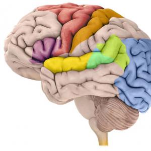
Rice. 7.1.Map of cytoarchitectonic fields of the human cerebral cortex (Brain Institute):
a- outside surface; b- internal; v- front; G- back surface. The numbers indicate the fields
of the brain, based on the evolutionary principle, employees of the Institute of the Brain of the USSR (founded in the 1920s in Moscow by O. Vogt, invited for this purpose) created detailed maps of the cytomyeloarchitectonic fields of the human brain (Fig. 7.1).
7.1. Zones and fields of the cerebral cortex
In the cerebral cortex, functional zones are distinguished, each of which includes several Brodmann fields(53 fields in total).
1st zone - motor - represented by the central gyrus and the frontal zone in front of it (4, 6, 8, 9 of Brodmann's field). When it is irritated, various motor reactions occur; when it is destroyed - impaired motor functions: weakness, paresis, paralysis (respectively, weakening, sharp decrease, disappearance
movements). In the motor zone, the areas responsible for the innervation of various muscle groups are represented differently. The zone involved in the innervation of the muscles of the lower limb is presented in the upper part of the 1st zone; muscles of the upper limb and head - in the lower part of the 1st zone. The largest area is occupied by the projection of facial muscles, muscles of the tongue and small muscles of the hand.
2nd zone - sensitive - Areas of the cerebral cortex posterior to the central sulcus (1, 2, 3, 5, 7 of Brodmann's field). When this zone is irritated, paresthesia occurs, when it is destroyed, the superficial and part of deep sensitivity fall out. In the upper sections of the postcentral gyrus, there are cortical centers of sensitivity for the lower extremity of the opposite side, in the middle sections - for the upper, and in the lower - for the face and head.
The 1st and 2nd zones are closely related to each other functionally. In the motor zone, there are many afferent neurons receiving impulses from proprioceptors - these are motosensory zones. There are many motor elements in the sensitive zone - these are sensorimotor zones that are responsible for the onset of pain.
3rd zone - visual - the occipital region of the cerebral cortex (17, 18, 19 Brodmann's fields). When field 17 is destroyed, loss of visual sensations (cortical blindness) occurs. Different parts of the retina are unequally projected in the 17th Brodmann field and have different locations. With the point destruction of the 17 field, the completeness of the visual perception of the environment is disturbed, since a portion of the field of view falls out. With the defeat of the 18th Brodmann field, the functions associated with the recognition of the visual image suffer, the perception of writing is impaired. When Brodmann's field 19 is damaged, various visual hallucinations occur, visual memory and other visual functions suffer.
4th zone - auditory - the temporal region of the cerebral cortex (22, 41, 42 Brodmann's fields). If 42 fields are hit, the sound recognition function is impaired. With the destruction of 22 fields, auditory hallucinations, impaired auditory orientational reactions, musical deafness occur. With the destruction of 41 fields - cortical deafness.
5th zone - olfactory - located in the pear-shaped gyrus (11 Brodmann's field).
6th zone - gustatory - 43 Brodmann's field.
7th zone - speech motor (according to Jackson - the center of speech) in right-handers is located in the left hemisphere. This zone is divided into 3 sections:
1) Broca's speech motor center (the center of speech praxis) is located in the posterior inferior part of the frontal gyri. He is responsible for the praxis of speech, i.e. ability to speak. It is important to understand the difference between Broca's center and the motor center of the speech-motor muscles (tongue, pharynx, face), which is located in the anterior central gyrus, posterior to Broca's zone. When the motor center of these muscles is damaged, their central paresis or paralysis develops. At the same time, a person is able to speak, the semantic side of speech does not suffer, but his speech is fuzzy, his voice is low-modulated, i.e. the quality of sound pronunciation is disturbed. When Broca's zone is affected, the muscles of the speech motor apparatus are intact, but the person is not able to speak like a child in the first months of life. This state is called motor aphasia;
2) Wernicke's sensory center located in the temporal zone. It is associated with the perception of oral speech. With his defeat, sensory aphasia occurs - a person does not understand oral speech (both someone else's and his own). Due to a lack of understanding of his own speech production, the patient's speech takes on the character of a "verbal salad", i.e. a set of unrelated words and sounds.
With a joint defeat of the Broca and Wernicke centers (for example, with a stroke, since they are both located in the same vascular basin), total (sensory and motor) aphasia develops;
3) center for the perception of written language is located in the visual area of the cerebral cortex - 18 Brodmann's field. With his defeat, agraphia develops - the inability to write.
There are similar, but undifferentiated zones in the subdominant right hemisphere, while the degree of their development is different for each individual. If the left-hander has a damaged right hemisphere, speech function is affected to a lesser extent.
At the macroscopic level, the cerebral cortex can be divided into sensory, motor and associative zones. Sensory (projection) zones, which include the primary somatosensory cortex, the primary zones of various analyzers (auditory, visual, gustatory, vestibular), have a connection with certain areas,
organs and systems of the human body, peripheral parts of the analyzers. The same somatotopic organization has also motor cortex. The projections of body parts and organs are presented in these areas according to the principle of functional significance.
Associative cortex, which includes the parietotemporal-occipital, prefrontal and limbic associative zones, is important for the implementation of the following integrative processes: higher sensory functions and speech, motor praxis, memory and emotional (affective) behavior. The associative parts of the cerebral cortex in humans are not only larger in their area than projection (sensory and motor) ones, but are also characterized by a thinner architectonic and neural structure.
7.2. The main types of higher mental functions and their disorders
7.2.1. Gnosis, types of agnosia
Gnosis (from the Greek. gnosis - cognition, knowledge) is the ability to cognize or recognize the surrounding world, in particular various objects of the surrounding world, using information coming from various cortical analyzers. At every moment of our life, analyzer systems supply the brain with information about the state of the external environment, about objects, sounds, smells around us, about the position of our body in space, which makes it possible for us to adequately perceive ourselves in relation to the world around us and correctly respond to all changes occurring around us.
Agnosia - These are disorders of recognition and cognition, reflecting violations of various types of perception (shape of an object, symbols, spatial relationships, speech sounds, etc.), arising from damage to the cerebral cortex.
Depending on the affected analyzer, visual, auditory and sensory agnosias are distinguished, each of which includes a large number of disorders.
Visual agnosias call such disorders of visual gnosis that arise when the cortical structures (and the nearest subcortical formations) of the posterior parts of the cerebral hemispheres (parietal and occipital regions) are damaged and proceed with the relative preservation of elementary visual functions (visual acuity, color perception, visual fields) [fields 18, 19 according to Brodman].
Object agnosia characterized by impaired visual recognition of objects. The patient can describe various features of the object (shape, size, etc.), but cannot recognize it. Using information from other analyzers (tactile, auditory), the patient can partially compensate for his defect, therefore such people often behave almost like blind - although they do not bump into objects, they constantly feel, sniff, listen. In milder cases, it is difficult for patients to recognize inverted, crossed out, superimposed images.
Optical-spatial agnosia occurs when the upper part of the parieto-occipital region is affected. The patient's orientation in space is disturbed. The right-left orientation suffers especially. Such patients do not understand the geographical map, do not orient themselves on the terrain, do not know how to draw.
Letter agnosia - violation of the recognition of letters, as a result, there is alexia.
Facial agnosia (prosopagnosia) - impaired recognition of faces, arising from the defeat of the posterior parts of the subdominant hemisphere.
Apperceptive agnosia characterized by the inability to recognize integral objects or their images while maintaining the perception of individual signs.
Associative agnosia - visual agnosia, characterized by a violation of the ability to recognize and name whole objects and their images while maintaining their distinct perception.
Simultaneous agnosia - inability to synthetically interpret groups of images that form a whole. It occurs with bilateral or right-sided lesions of the occipital-parietal regions of the brain. The patient cannot simultaneously perceive several visual objects or situations in general. Only one object is perceived, more precisely, only one operational unit of visual information is processed, which is currently the object of the patient's attention.
Auditory agnosias are divided into violations of speech phonemic hearing, intonation side of speech and non-speech auditory gnosis.
Auditory agnosias associated with phonemic hearing, occur mainly with damage to the temporal lobe of the dominant hemisphere. Due to impairment of phonemic hearing, the ability to distinguish the sounds of speech is lost.
Auditory non-verbal (simple) agnosia occurs when the cortical level of the auditory system of the right hemisphere (nuclear zone) is damaged; the patient is not able to determine the meanings of various everyday (object) sounds, noises. Sounds such as the creak of a door, the noise of water, the clink of dishes cease to be carriers of a certain meaning for these patients, although hearing as such remains intact, and they can distinguish sounds by height, intensity, timbre. With damage to the temporal region, a symptom such as arrhythmia. Patients cannot correctly assess by ear various rhythmic structures (a series of claps, taps) and cannot reproduce them.
Amusia- auditory agnosia with impaired musical abilities that the patient had in the past. Motor amusia is manifested by the impossibility of playing familiar melodies; sensory- violation of recognition of familiar melodies.
Violation of the intonation side of speech occurs when the temporal region of the subdominant hemisphere is damaged, while the perception of the emotional characteristics of the voice, the distinction between male and female voices is lost, and one's own speech loses its expressiveness. Such patients cannot sing.
Sensitive agnosias are expressed in the lack of recognition of objects when they act on the receptors of superficial and deep sensitivity.
Tactile agnosia, or astereognosis occurs when the post-central areas of the cortex of the lower parietal region are affected, bordering on the zones of representation of the hand and face in the 3rd field, and is manifested by the inability to perceive objects by touch. Tactile perception is preserved, therefore, the patient, feeling the object with closed eyes, describes all its properties (“soft”, “warm”, “prickly”), but cannot identify this object. Sometimes difficulties arise in identifying the material from which the object is made. This type of violation is called tactile agnosia of object texture.
Finger agnosia, or Terstmann's syndrome observed when the inferior parietal cortex is affected, when the ability to call the fingers on the hand contralateral to the lesion is lost with closed eyes.
Disorders of the "body scheme", or autopagnosia occurs when the upper parietal region of the cerebral cortex is damaged, which is adjacent to the
vic sensory cortex of the skin-kinesthetic analyzer. Most often, the patient has impaired perception of the left half of the body due to damage to the right parietal region of the brain. The patient ignores the left limbs, the perception of his own defect is often impaired - anosognosia (Anton-Babinsky syndrome), those. the patient does not notice paralysis, sensory disturbances in the left extremities. In this case, false somatic images may appear in the form of a feeling of "someone else's hand", doubling of limbs - pseudopolymelia, increase, decrease in body parts, pseudoamelia -"Absence" of a limb.
7.2.2. Praxis, types of apraxia
Praxis (from the Greek. praxis - action) - the ability of a person to perform appropriate sequential complexes of movements and to perform purposeful actions according to the developed plan.
Apraxia - Praxis disorders, which are characterized by a loss of skills developed in the process of individual experience, complex purposeful actions (everyday, industrial, symbolic gestures) without pronounced signs of central paresis or impaired coordination of movements.
According to the classification proposed by A.R. Luria, there are 4 forms of apraxia.
Kinesthetic apraxia occurs with damage to the lower parts of the postcentral gyrus of the cerebral cortex (fields 1, 2, partly 40, mainly of the left hemisphere). In these cases, there are no clear movement disorders, muscle paresis, but movement control is impaired. Patients can hardly write, the accuracy of reproduction of hand poses is impaired (posture apraxia), they cannot depict this or that action without an object (smoking a cigarette, combing). Partial compensation for this disorder is possible with increased visual control over the execution of movements.
With spatial apraxia the correlation of one's own movements with space is violated, spatial representations "up-down", "right-left" are violated. The patient cannot give a straightened hand a horizontal, frontal, sagittal position, draw an image oriented in space, while writing errors occur in the form of "mirror writing". Such a violation occurs when the parieto-occipital parts of the cortex are affected at the border of the 19 and 39 fields, bilateral or isolated of the left hemisphere. It
often combined with visual optic-spatial agnosia; in this case, a complex picture of apractoagnosis arises. This type of disorder also includes constructive apraxia - the difficulty of constructing a whole from individual objects (Koos cubes, etc.).
Kinetic apraxia associated with damage to the lower parts of the premotor cortex (6 and 8 fields). In this condition, there is a violation of the temporary organization of movements (automation of movements). This form of apraxia is characterized by motor perseverations, manifested in the uncontrolled continuation of the once begun movement. It is difficult for the patient to switch from one elementary movement to another, as if he is stuck on each of them. This is especially pronounced when writing, drawing, performing graphic tests. Often, hand apraxia is combined with speech disorders (motor efferent aphasia), and a commonality of mechanisms underlying the pathogenesis of these conditions has been established.
Regulatory(or prefrontal) form of apraxia occurs when the convexital prefrontal cortex is damaged in front of the premotor parts of the frontal lobes and is manifested by a violation of the programming of movements. Disabled conscious control over their implementation, the necessary movements are replaced by patterns and stereotypes. Perseverations are characteristic, but already systemic, i.e. not elements of the motor program, but the entire program as a whole. If such patients are offered to write something under dictation, and after executing this command, they are asked to draw a triangle, then they will circle the outline of the triangle with movements characteristic of writing. With a gross disintegration of voluntary regulation of movements in patients, symptoms of echopraxia are observed in the form of imitative repetitions of the doctor's movements. This type of violation is closely related to the violation of speech regulation of motor acts.
7.2.3. Speech. Types of aphasia
Speech is a specific human mental function that can be defined as the process of communication through language. Allocate impressive speech(perception of oral, written speech, its decoding, awareness of meaning and correlation with previous experience) and expressive speech(begins with the intention of the utterance, then goes through the stage of internal speech and ends with a detailed external speech utterance).
Aphasia - complete or partial speech impairment that occurs after the period of its normal formation, due to local
lesion of the cortex (and adjacent subcortical formations) of the dominant cerebral hemisphere. Aphasias are manifested in the form of violations of the phonemic, morphological and syntactic structure of one's own speech and understanding of the addressed speech, while the movements of the speech apparatus, providing articulate pronunciation, and elementary forms of hearing are preserved.
Sensory aphasia (acoustic-gnostic aphasia) occurs when the posterior third of the temporal gyrus is affected (field 22); was first described by K. Wernicke in 1864. It is characterized by the impossibility of normal perception of both someone else's and his own oral speech. It is based on a violation of phonemic hearing, i.e. loss of the ability to distinguish the sound composition of words (distinguishing between phonemes). In Russian, phonemes are all vowels and their stress, as well as consonants and their voiced-voicelessness, hardness-softness. In the case of incomplete destruction of the zone, it is difficult to perceive fast or "noisy" speech (for example, when two or more interlocutors speak). In addition, patients practically cannot distinguish words that are similar in sound, but different in meaning: "ear-voice-single" or "fence-cathedral".
In more severe cases, the person's ability to perceive the phonemes of their native language completely disappears. Patients do not understand the speech addressed to them, perceiving it as noise, conversation in an unknown language. There is a secondary decay and active spontaneous oral speech, since there is no auditory control, i.e. understanding and assessment of the correctness of the spoken words. Speech utterances are replaced by the so-called "verbal salad", when patients pronounce words and expressions incomprehensible in their sound composition. Sometimes it is possible to pronounce familiar words, however, in them, patients often replace some sounds with others; such a violation is called literal paraphasias. When replacing whole words, they talk about verbal paraphasias. In such patients, writing under dictation is disturbed, the repetition of the words heard, reading aloud is sharply difficult. However, ear for music with a given localization of the pathological focus is usually not impaired and articulation is completely preserved.
At motor aphasia (speech apraxia) there are violations of the pronunciation of words with a relative safety of speech perception.
Afferent motor aphasia occurs when the lower parts of the postcentral parts of the parietal region of the brain are damaged. Such patients often cannot arbitrarily make various sounds, not
can puff out one cheek, stick out their tongue, lick their lips. Sometimes control only of complex articulatory movements suffers (difficulty in pronouncing words like "propeller", "space", "sidewalk"), however, at the same time, patients feel mistakes in pronunciation, but are not able to correct them, since "their mouth does not obey ". Violation of articulation also affects written speech in the form of replacing letters with similar ones in pronunciation.
Efferent motor aphasia (Broca's classical aphasia, fields 44, 45) occurs when the lower parts of the premotor cortex (posterior third of the inferior frontal gyrus) of the dominant hemisphere are destroyed. The leading defect in this violation is a partial or complete loss of the ability to smoothly switch motor impulses in time. Violations of arbitrary simple movements of the lips, tongue with this pathology are not observed. Such patients can pronounce individual sounds or syllables, but cannot combine them into words, phrases. In this case, pathological inertness of articulatory actions arises, which manifests itself in the form speech perseverations(constant repetition of the same syllable, word or expression). Often this verbal stereotype ("embolus") becomes a substitute for all other words. In erased cases, difficulties arise when pronouncing words or expressions that are "difficult" in a motor sense. Due to the defeat of connections with various "speech zones", violations of writing, reading and even understanding of speech can also occur.
Dynamic motor aphasia occurs when the prefrontal divisions are damaged (9, 10, 46 fields). In this case, the sequential organization of speech utterance is disrupted, active productive speech is disrupted, and reproductive (repeated, automated) is preserved. The patient can repeat the phrase, but he cannot construct a statement on his own. Passive speech is possible - monosyllabic answers to questions, often echolalia (repetition of the interlocutor's word).
With the defeat of the lower and posterior parts of the parietal and temporal regions, development of amnestic aphasia (on the border of 37 and 22 fields). This disorder is based on the weakness of visual representations, visual images of words. This type of violation is also called nominative amnestic aphasia, or opticomnestic aphasia. Patients repeat words well and speak fluently, but cannot name objects. The patient easily recalls the purpose of objects (pen - "what they write"), but they cannot remember their names. A doctor's advice will often make the assignment easier,
since the understanding of speech remains intact. Patients are capable of dictation and reading, while spontaneous writing is impaired.
Acoustic-mnestic aphasia occurs when the middle parts of the temporal region of the dominant hemisphere, located outside the zone of the sound analyzer, are affected. The patient correctly understands the sounds of the native language, the addressed speech, but is unable to memorize even a relatively small text due to a gross impairment of auditory memory. The speech of these patients is characterized by scarcity, frequent omission of words (more often nouns). Prompts when trying to reproduce words do not help such patients, since speech traces are not retained in memory.
Semantic aphasia occurs when the cortical fields 39 and 40 of the parietal lobe of the left hemisphere are affected. The patient does not understand speech formulations reflecting spatial relationships. So, the patient cannot cope with tasks, for example, draw a circle under a square, a triangle over a line, not understanding how to position the figures relative to each other; the patient does not understand, cannot understand the comparative constructions: “Sonya is lighter than Mani, and Manya is lighter than Olya; which of them is the lightest, the darkest? " The patient does not catch the change in the meaning of the phrase when the word is rearranged, for example: "Students stood at the showcase with books", "Students stood at the showcase with books." It is not possible to understand the attributive constructions: are the father of the brother and the brother of the father the same person? The patient does not understand proverbs and metaphors.
Aphasias should be distinguished from other speech disorders arising from brain damage or functional disorders, such as dysarthria, dyslalia.
Dysarthria - a complex concept that unites such speech disorders in which not only pronunciation suffers, but also tempo, expressiveness, fluency, modulation, voice and breathing. This disorder can be caused by central or peripheral paralysis of the muscles of the speech-motor apparatus, damage to the cerebellum, striopallidal system. In this case, speech, reading and writing impairments most often do not occur. Distinguish between cerebellar, pallidary, striatal and bulbar dysarthria.
A speech disorder associated with a violation of sound pronunciation is called dislalia. It occurs, as a rule, in childhood (children "do not pronounce" certain sounds) and lends itself to speech therapy correction.
Alexia (from the Greek. a- denied. particle and lexis- word) - a violation of the process of reading or mastering it with the defeat of various parts of the cortex of the dominant hemisphere (fields 39-40 according to Brodman). There are several forms of alexia. When the cortex of the occipital lobes is damaged due to a violation of the processes of visual perception in the brain, optical alexia, in which either letters (literal optical alexia) or whole words (verbal optical alexia) are not defined. With unilateral optical alexia, lesions of the occipital-parietal parts of the right hemisphere, half of the text (usually the left) is ignored, while the patient does not notice his defect. Due to impaired phonemic hearing and sound-letter analysis of words, auditory (temporal) alexia as one of the manifestations of sensory aphasia. The defeat of the lower parts of the premotor region of the cortex leads to disruption of the kinetic organization of the speech act and the emergence kinetic (efferent) motor alexia, included in the structure of the syndrome of efferent motor aphasia. When the cortex of the frontal lobes of the brain is damaged, regulatory mechanisms are disturbed and a special form of alexia arises in the form of a violation of the purposeful nature of reading, disconnection of attention, and its pathological inertia.
Agrafia (from the Greek. a- denied. particle and grapho- I write) - a violation characterized by the loss of the ability to write with sufficient preservation of intelligence and formed writing skills (field 9 according to Brodman). It can manifest itself as a complete loss of writing ability, gross distortion of the spelling of words, gaps, inability to connect letters and syllables. Aphatic agraphia occurs with aphasia and is caused by defects in phonemic hearing and auditory-speech memory. Apractical agraphia occurs with ideational aphasia, constructive- with constructive aphasia. Also stands out pure agraphia, not associated with other syndromes and due to the defeat of the posterior parts of the second frontal gyrus of the dominant hemisphere.
Akalculia (from the Greek. a- denied. particle and lat. calculatio- counting, calculation) is described by S.E. Henschen in 1919. Characterized by violation of counting operations (fields 39-40 according to Brodmann). Primary acalculia as a symptom that does not depend on other disorders of higher mental functions, it is observed with damage to the parieto-occipital-temporal cortex of the dominant hemisphere and represents a violation of understanding spatial relationships, difficulty in performing digital operations with the transition through
a dozen associated with the bit structure of numbers, the inability to distinguish between arithmetic signs. Secondary acalculia can occur with damage to the temporal divisions due to a violation of the oral count, the occipital divisions due to the non-discrimination of similarly written numbers, the prefrontal divisions due to a violation of purposeful activity, planning and control of counting operations.
7.3. Features of the development of speech function in children in health and disease
Normally, children acquire the ability to speak and understand speech addressed to them during the first 3 years of life. In the 1st year of life, speech develops from the so-called humming to pronouncing syllables or simple words. In the 2nd year of life, a gradual accumulation of vocabulary occurs, and at about 18 months, children first begin to pronounce combinations of two words related in meaning. This stage is a harbinger of the development of complex grammar rules by children, which, according to some linguists, are a basic characteristic of human languages. In the 3rd year, the child's vocabulary increases from ten to hundreds of words, the structure of sentences becomes more complicated - from phrases consisting of two words to complex sentences. By the age of 4, children have practically mastered all the basic rules of the language. The development of expressive speech lags slightly behind the impressive one. Pronunciation of legible words requires accurate distinction between speech sounds and perfect functioning of the motor systems under the control of hearing. The clear pronunciation of all phonemes of the language improves over the years and not all children master it by the time they reach school age. Individual inaccuracies in the pronunciation of some consonants, which generally do not reduce speech intelligibility, are considered more a sign of brain immaturity than speech disorders.
If a child with normal intelligence and hearing is damaged in the speech areas of the cerebral hemispheres as a result of trauma or brain diseases in the first 3 years of life, then alalia - absence or underdevelopment of speech. Alalia, like aphasia, can be divided into motor and sensory.
Alalia may be a clinical manifestation of a complex impairment of speech function, which is called general speech underdevelopment(a form of speech pathology in children with normal hearing and primary intact intelligence, when the formation of all components of the speech system is disturbed).
7.4. Memory
In the most general sense, memory is called the retention of information about the stimulus after its action has already ceased. There are four phases of memory processes: fixation, storage, reading and reproduction of a trace.
In terms of duration, memory processes are divided into three categories:
1. Instant memory- short-term imprinting of traces, lasting a few seconds.
2. Short-term memory- imprinting processes that last several minutes.
3. Long term memory- long (possibly throughout life) preservation of traces of memory (dates, events, names, etc.).
In addition, memory processes can be characterized in terms of their modality, i.e. type of analyzer systems. Accordingly, visual, auditory, tactile, motor, olfactory memory is distinguished. There is also an affective or emotional memory, or memory for emotionally charged events. Various areas of the brain were identified that are responsible for one or another type of memory (hippocampus, cingulate gyrus, anterior nuclei of the thalamus, mammillary bodies, septa, vault, amygdala complex, hypothalamus), but, by and large, memory, like any complex mental process, is associated with the work of the whole brain, therefore, it is possible to talk about memory centers only conditionally.
Memory impairments are of various types, and the literature describes cases of not only weakening (hypomnesia) or complete loss of memory (amnesia), but also its pathological increase (hypermnesia).
Hypomnesia, or memory impairment, can be of various origins. It can be associated with age-related changes, brain diseases, or be congenital. Such patients, as a rule, are characterized by the weakening of all types of memory. Memory impairment with the loss of the ability to retain and reproduce acquired knowledge is called amnesia.
With damage at the level of the limbic system, the so-called Korsakov syndrome. Patients with Korsakov's syndrome have practically no memory for current events, for example, they greet the doctor several times, cannot remember what they did a few minutes ago, at the same time, these
patients with relatively well preserved traces of long-term memory, they are able to remember the events of the distant past.
Similar conditions can occur with transient hypoxia of the brain, some intoxications (for example, with carbon monoxide poisoning). This memory impairment is also called fixative amnesia. With a pronounced violation of memorizing new facts and circumstances, amnestic disorientation develops in time, in the space of one's own personality. Another example of a kind of temporary violation of all types of memory is global transient amnesia with transient ischemia in the vertebrobasilar basin.
A special group of memory impairments are the so-called pseudoamnesia(false memories), characteristic of patients with massive lesions of the frontal lobes of the brain. The problems of memorizing the material are associated in this case with a violation not so much of the memory itself, but of purposeful memorization, since in these patients the process of forming intentions, plans, behavior programs is grossly violated, i.e. the structure of any conscious mental activity suffers.
7.5. Syndromes of lesion of the cerebral cortex
Syndromes of damage to the cerebral cortex include symptoms of loss of function or irritation of the cortical centers of various analyzers (Table 13).
Table 13.Syndromes of lesion of the cerebral cortex Frontal Lobe Syndromes


7.6. Violation of HMF in cerebellar lesions
Violation of the HMF in lesions of the cerebellum is explained by the loss of its coordinating role in relation to various parts of the cerebellum. Cognitive disorders develop in the form of impaired working memory, attention, planning and control of actions, i.e. disorder of the sequence of actions. Also, there are visual-spatial disturbances, acoustic-mnestic aphasia, difficulties in counting, reading and writing, and even facial agnosia.
Corpus callosum syndrome accompanied by mental disorders in the form of confusion, progressive dementia. Amnesia and confabulations (false memories), a feeling of "already seen", congestion, apraxia, akinesia are noted. The orientation in space is violated.
Frontal-callous syndrome characterized by akinesia, amimia, astasia-abasia, spontaneity, reflexes of oral automatism, memory impairments, decreased criticism of one's condition, grasping reflexes, apraxia, Korsakov's syndrome, dementia.
A rotating globe and a "patchwork quilt" of the countries depicted on it - such a map helps us understand exactly where we are, as well as the fact that states and peoples differ from each other and have very specific boundaries. Now a similar map has appeared for the outer layer of the brain - its cortex, on which each hemisphere was divided into 180 separate"Countries"... Moreover, ninety-seven areas of them have never been described before, despite the obvious differences in structure, function and significant connection with neighbors. New brain map published inNature.
Illustration: M. F. Glasser, D. C. Van Essen
“Each individual area on the map contains cells with similar structures, functions and relationships. But these "regions" differ from each other like different countries and have well-defined boundaries, as well as a unique culture ",
Commented by David Van Essen, a neurologist at the University of Washington School of Medicine in St. Louis, Missouri, who oversaw the study.
Neuroscientists have long sought to divide the brain into smaller pieces to better understand how it works as a whole. One of the most famous brain maps divided the cortex into 52 regions based on the different arrangement of cells in tissue (now known as Brodmann's fields). More recent maps have relied on magnetic resonance imaging (MRI) data - for example, functional MRI, which measures blood flow in response to various mental tasks.
However, so far, most of these "cartographic" studies have been done with just one type of measurement, which, as noted by Thomas Yeo, a neuroscientist at the National University of Singapore, not only does not provide a complete picture of how the whole brain works, but it can mislead researchers. The new map is based on multiple MRI measurements that maximize focus on cortical areas and therefore help assess them best.
Divide and rule
To build the map, a team led by neuroscientist Mathew Glasser of the University of Washington School of Medicine in St. Louis used images from 210 healthy young people who participated in the Human Connectome project to map brain structures and how they function. ... The study included information on the thickness of the cortex, brain function, axonal connections between regions, the topographic organization of cells in tissue, and the content of myelin, a substance that is responsible for electrical insulation of axons.
Glasser zoned the cerebral cortex on the basis of significant changes in two or more properties, which he used to "set" boundaries on the map. Data processing was carried out using machine learning algorithms.
“If you slowly“ crawl ”along the surface of the bark, then at some point you will find a place where properties begin to change, or even one where several independent changes are found in the same place,”
The researcher noted.
The technique confirmed the presence of 83 previously known brain regions and identified another 97 new ones. Scientists have checked the map by observing the work of these regions in 210 people. They found that the map was accurate enough, but the size of its zones varied slightly from person to person. These differences will help to understand in a new way the individual variability of cognitive abilities, as well as identify the risks of developing diseases.
The edge of the achievable
"While the focus of this work has been to create a beautiful and average brain template, it does open up the opportunity for further exploration of unique combinations of individual traits with intellectual and creative abilities, that is, what makes each of us unique."
This is Rex Jung, a neuropsychologist at the University of New Mexico at Albuquerque.
But with all the obvious pluses, there are also minuses: the card is limited in several important aspects. Most importantly, it tells very little about the biochemical basis of the brain or about the activity of individual neurons, their small groups.
“It looks like a fantastic Google Earth map that can even showcase your area of a city or even your backyard. However, you cannot consider how your neighbors are moving, where they are going to move and what kind of work they have. "
Jung speaking.
Glasser expects this project to become version 1.0.
“This does not mean that this version is final, but what we got now is much better than what we had before,”
The scientist remarks reasonably.
A multi-modal parcellation of human cerebral cortex
Matthew F. Glasser, Timothy S. Coalson, Emma C. Robinson, David C. Van Essen et all.
Nature (2016) doi: 10.1038 / nature18933
The cerebral cortex plays the role of capturing, receiving and transmitting various types of stimuli. They transmit pulses to other analyzer cores.
Ivan Petrovich Pavlov described the cerebral hemispheres as an accumulation of cortical analyzers, where responses are generated that affect the activity of the body.
Study history
In Germany, in 1909, physician-scientist Corbinian Brodmann was the first to build and describe maps of Brodmann's cytoarchitectonic fields. Ten years later, O. Vogt and C. Vogt studied and described more than a hundred myelo-architectonic regions in the cerebral hemispheres.
As a result, I.N. Filimonov and S.A. Sarkisov released a Brodman map of the cerebral hemispheres, which describes 47 fields.
Brodmann's fields are most often used to study the neural organization of the brain and its functioning. The division of a particular area of the brain to a specific section of the fields was carried out on the basis of histological analysis - color staining according to Nissl.
Groups of fields of the brain according to Brodman
Description of Brodmann's fields in the cerebral cortex by zones:
- The first zone is motor responsible for the reactions of active movements. This zone includes 4, 6, 8, 9 Brodmann fields. 4 is responsible for motor skills, located in the precentral. 6 stands out in the anterior regions of the precentral gyrus and in the region of the middle frontal gyrus. 8 coordinates the voluntary mobility of the eyes and is located in the posterior sections of the superior and middle frontal gyri. 9 is located in the prefrontal region.
- The second zone is affective... Includes the areas of the cerebral cortex behind the Roland groove. There are 1, 2, 3, 5 and 7 fields here. The upper part of the zone is responsible for the tactile sensations of the legs and genitals. Underlying areas - for sensations in the hands, skull and mouth. The second zone directly interacts with the first. The first zone contains afferent nerve cells that receive stimuli from - these are motor sensory areas. The second zone contains the motor components - these are the sensorimotor areas that regulate the formation and intensity of pain feelings.
- The third zone is the visual, located in the occipital region of the cerebral cortex. It includes 17, 18, 19 Brodman fields. 17th belong to the primary visual area, and 18 and 19 - the secondary visual area. From the secondary areas, visual stimuli enter the primary areas and are already processed there. With pathologies of the 17th Brodmann field, cortical blindness occurs - a loss of visual perception. In case of violation of 18, the function of identification and perception of written speech is affected. With pathologies 19, hallucinations and violations of figurative memory are noted.
- The fourth zone is the auditory, allocated in the temporal area of the cerebral cortex. It includes 22, 41, 42, 52 fields. With the defeat of 22, auditory hallucinations are noted, orientation to sound suffers, and musical deafness occurs. With pathologies 42, sound recognition suffers, and damage 41 - cortical deafness, that is, a complete loss of auditory perception. 52 is a zone that is responsible for the spatial perception of voices, sounds and speech.
- In the fifth - the olfactory zone includes 11 and 29 fields, which stand out in the pear-shaped gyrus. Responsible for recognizing various odors.
- Sixth zone - gustatory, including the 43rd field.
- Seventh zone - speech, in right-handers it is located in the left hemisphere. This includes 22 fields -, 37 - controls voluntary speech and its comprehension, 47 - areas of singing, 44 and 45 - Brokk's speech centers.
- 24, 25 and 26 fields Brodmann perform the task of recognizing mismatches and errors.
Brodmann Fields Scheme:

Centers of the first signaling system
The first signaling system is found in both Homo sapiens and other living beings. It performs the functions of understanding and analyzing irritations emanating from the surrounding world and manifested in the form of sensations and representations.
The centers of the first signaling system are located in the nuclei of sensory sensitivity analyzers, motor analyzers, auditory, skin, visual and olfactory analyzers.
They are indicated in the superior and inferior parietal regions of the brain, in the precentral and supra-marginal sulcus, as well as in the thickness of the lateral sulcus (auditory nuclei).
The purpose of this system is presented in the Brodmann field table:
| Brodman fields | Analyzer kernels | Functions |
|---|---|---|
| 1, 2, 3, 5, 7 | Cortical | Responsible for the perception of temperature and pain, as well as tactile sensations. The affective pathways stretching to the cerebral cortex intersect in the region and the medulla oblongata. Because of this, the functions of any of the hemispheres are controlled by the diametrically inverse part of the body. |
| 6 and 4 | Motor | Here, neurons are released, the reactions from which are controlled by the muscles of the lower body and facial muscles. |
| 8 | Skulls and eyes | The nuclei that control the movements of the head and eyes. |
| 40 | Motor | Control voluntary purposeful movements of an animal or a person. |
| 18, 17 and 19 | Spotting | They are responsible for visual memory, perception of images and orientation in unfamiliar places. |
| 7 | Dermal | Performs tactile functions of recognizing objects and surfaces to the touch and other types of skin sensitivity. |
| 41, 42 and 52 | Auditory | Perception and memorization of sounds from the outside. |
| 43 | Olfactory | Phylogenetically, the oldest region of the cerebral cortex. Provides the function of smelling and memorizing. Closely related to the sense of taste. |
The picture shows the fields of the cerebral hemispheres according to Brodman and their functions:

Second signaling system
The second signaling system exists exclusively in Homo sapiens; its emergence is explained by speech abilities and thinking.
The second signaling system is located in the nuclei of the motor analyzer of the written text, the speech-motor analyzer, the understanding of verbal and non-verbal speech. The nuclei of the second system are spread out in the posterior and central regions of the underlying frontal gyrus, as well as in the overlying temporal sulcus and below the parietal lobe.
The cores determine the purpose of the second signaling system.
The core of the perception of writing includes a field 40, it is responsible for the image and understanding of the written letters. It also controls hand movements, turns of the skull and eyes, fine motor skills.
The speech-motor area consists of field 44 - this is Brocca's speech center, as well as 45, associated with musical perception. This nucleus is interconnected with the areas of movement, since the motility of many muscles of the tongue and mouth plays a role in the speech process. Performs the functions of oral reproduction of sounds, words and phrases.
The auditory core of oral speech consists of fields 42 and 22, controls the task of recognizing and understanding oral speech sounds.
The core of written speech - field 29, provides the functions of analysis, perception and understanding of the written text and reading.
In the cerebral cortex, zones are distinguished- Brodmann's fields (German physiologist).
1st zone- motor - represented by the central gyrus and the frontal zone in front of it - 4, 6, 8, 9 of Brodmann's field. With her irritation - various motor reactions; with its destruction - violations of motor functions: weakness, paresis, paralysis (respectively - weakening, sharp decrease, disappearance).
In the 50s of the twentieth century. have established that in the motor zone, different muscle groups are represented differently. The muscles of the lower limb are in the upper section of the 1st zone. The muscles of the upper limb and head are in the lower part of the 1st zone. The largest area is occupied by the projection of facial muscles, muscles of the tongue and small muscles of the hand.
2nd zone- sensitive - areas of the cerebral cortex posterior to the central sulcus (1, 2, 3, 4, 5, 7 of Brodmann's field). When this zone is irritated, sensations arise, when it is destroyed - loss of skin, proprio, intersensitivity. Hyposthesia - decreased sensitivity, anesthesia - loss of sensitivity, paresthesia - unusual sensations (goose bumps). Upper sections of the zone - the skin of the lower extremities, genitals is presented. In the lower sections - the skin of the upper limbs, head, mouth.
The 1st and 2nd zones are closely related to each other functionally. There are many afferent neurons in the motor zone that receive impulses from proprioceptors - these are motosensory zones. In the sensitive zone, many motor elements - these are sensorimotor zones - are responsible for the onset of pain.
3rd zone- the visual zone - the occipital region of the cerebral cortex (17, 18, 19 Brodmann's fields). With the destruction of the 17th field - loss of visual sensations (cortical blindness).
Different areas of the retina are unequally projected in the 17th Brodmann field and have a different arrangement with the point destruction of the 17th field, the vision of the environment is lost, which is projected onto the corresponding areas of the retina. With the defeat of 18 Brodmann's field, the functions associated with the recognition of the visual image suffer and the perception of writing is impaired. With the defeat of Brodmann's field 19, various visual hallucinations occur, visual memory and other visual functions suffer.
4th - auditory zone- the temporal region of the cerebral cortex (22, 41, 42 Brodmann's fields). If 42 fields are damaged, the sound recognition function is impaired. When the 22 fields are destroyed, auditory hallucinations, impaired auditory orientation reactions, musical deafness occur. With the destruction of 41 fields - cortical deafness.
5th zone- olfactory - located in the pear-shaped gyrus (11 Brodmann's field).
6th zone- gustatory - 43 Brodman field.
7th zone- the speech motor zone (according to Jackson - the center of speech) - in most people (right-handed people) is located in the left hemisphere.
This zone consists of 3 sections.
Broca's propulsion center- located in the lower part of the frontal gyrus - this is the motor center of the muscles of the tongue. With the defeat of this area - motor aphasia.
Wernicke's sensory center- located in the temporal zone - associated with the perception of oral speech. With a defeat, sensory aphasia occurs - a person does not perceive spoken language, pronunciation suffers, as well as the perception of his own speech is impaired.
Written Speech Perception Center- located in the visual area of the cerebral cortex - 18 Brodmann's field, similar centers, but less developed, are also in the right hemisphere, the degree of their development depends on the blood supply. If the left-hander has a damaged right hemisphere, speech function is affected to a lesser extent. If the left hemisphere is damaged in children, then the right hemisphere takes over its function. In adults, the ability of the right hemisphere to reproduce speech functions is lost.
There are 53 fields in total (according to Brodman).