Biology drawings in animal life. Biology is the science of life
What is biology? Biology is the science of life, of living organisms that live on Earth.
Picture 3 from the presentation "Science" to biology lessons on the topic "Biology"
Dimensions: 720 x 540 pixels, format: jpg. To download a picture for free biology lesson, right-click on the image and click "Save Image As ...". To show pictures in the lesson, you can also download the entire presentation "Science.ppt" with all pictures in a zip archive for free. The archive size is 471 KB.
Download presentationBiology
"Research methods in biology" - The history of the development of biology as a science. Experiment planning, choice of technique. Lesson plan: For solving what global problems of mankind is knowledge of biology necessary? Topic: Frontier disciplines: Task: Morphology anatomy physiology systematics paleontology. The Significance of Biology ". Biology is a nuka about life.
"Scientist Lomonosov" - He emphasized the importance of exploring the Northern Sea Route and the development of Siberia. November 19, 1711 - April 15, 1765 (53 years old). June 10, 1741. Discoveries. Developed atomic-molecular concepts of the structure of matter. Ideas. Eliminated phlogiston as a chemical agent. Job. Being a supporter of deism, he considered natural phenomena materialistically.
"Botanist Vavilov" - All-Union Institute of Applied Botany. In 1906 Vavilov Nikolai Ivanovich. In 1924 Completed by: Babicheva Roksana and Zhdanova Lyudmila, pupils of grade 10 B. Vavilov's authority as a scientist and organizer of science grew. In Merton (England), in the genetic laboratory of the Horticultural Institute. N.I. Vavilov was born on November 26, 1887 in Moscow.
"Project activity" - E.V. Alekseeva Lecture plan. The teacher becomes the author of the project. Browse additional resources. Technologization of the information model of the educational process. Designing a biology lesson. Project activities... Theory and practice. (Project method). Stages of the teacher's work. Theory and practice. The main blocks in projects.
"Science of Wildlife" - Design of workbooks. 3. Biology is the science of living nature. Biology is the science of living nature. Bacteria. Mushrooms. They consist of one cell and do not have a nucleus. Mark Cicero. Biology studies living organisms. Have chlorophyll and form in the light organic matterreleasing oxygen. Question: What does biology study?
The life sciences are moving from large to small. More recently, biology described exclusively the external features of animals, plants, bacteria. Molecular biology studies living organisms at the level of interactions between individual molecules. Structural biology - examines processes in cells at the atomic level. If you want to learn how to "see" individual atoms, how structural biology works and "lives" and what devices it uses, you are here!
The general partner of the cycle is the company: the largest supplier of equipment, reagents and consumables for biological research and production.
One of the main missions of "Biomolecule" is to get to the very roots. We do not just tell what new facts the researchers discovered - we talk about how they discovered them, we try to explain the principles of biological methods. How to get a gene out of one organism and insert it into another? How can you trace the fate of several tiny molecules in a huge cell? How do you fire one tiny group of neurons in a huge brain?
And so we decided to talk about laboratory methods in a more systematic way, to bring together the most important, most modern biological methods in one heading. To make it more interesting and clearer, we thickly illustrated articles and even added animations here and there. We want the articles of the new section to be interesting and understandable even to a casual passer-by. And on the other hand, so that they are so detailed that even a professional could discover something new in them. We have collected the techniques in 12 large groups and are going to make a biomedical calendar based on them. Wait for updates!
Why Structural Biology?
As you know, biology is the science of life. She appeared in the very early XIX century and the first hundred years of its existence was purely descriptive. The main task of biology at that time was considered to be to find and characterize as many species of various living organisms as possible, and a little later - to identify the relationship between them. Over time and with the development of other fields of science, several branches with the prefix "molecular" emerged from biology: molecular genetics, molecular biology and biochemistry - sciences that study living things at the level of individual molecules, and not by the appearance of the organism or the interposition of its internal organs. Finally, quite recently (in the 50s of the last century) such a field of knowledge as structural biology - a science that studies processes in living organisms at the level of change spatial structure individual macromolecules. In fact, structural biology is at the intersection of three different sciences. Firstly, this is biology, because science studies living objects, secondly, physics, since the widest arsenal of physical experimental methods is used, and thirdly, chemistry, since changing the structure of molecules is the object of this particular discipline.
Structural biology studies two main classes of compounds - proteins (the main "working body" of all known organisms) and nucleic acids (the main "information" molecules). It is thanks to structural biology that we know that DNA has a double helix structure, that tRNA should be depicted as a vintage letter "G", and in the ribosome there are large and small subunits consisting of proteins and RNA in a certain conformation.
Global goal structural biology, like any other science - "to understand how everything works." In what form is the protein chain coiled, which causes cells to divide, how the packaging of the enzyme changes during the chemical process that it carries out, where the growth hormone and its receptor interact - these are the questions this science answers. Moreover, a separate goal is to accumulate such a volume of data so that these questions (for a still unexplored object) can be answered on a computer without resorting to an expensive experiment.

For example, you need to understand how the bioluminescence system works in worms or fungi - they decoded the genome, based on these data, they found the required protein and predicted its spatial structure along with the mechanism of operation. It is worth recognizing, however, that while such methods exist only in their embryonic stage, it is still impossible to accurately predict the structure of a protein, having only its gene. On the other hand, the results of structural biology have applications in medicine. Many researchers hope that knowledge about the structure of biomolecules and the mechanisms of their work will allow the development of new drugs on a rational basis, and not by trial and error (high-throughput screening, strictly speaking), as is done most often now. And this is not science fiction: there are already many drugs created or optimized using structural biology.
History of structural biology
The history of structural biology (Fig. 1) is rather short and starts in the early 1950s, when James Watson and Francis Crick, based on the data of Rosalind Franklin on X-ray diffraction on DNA crystals, assembled a model of the now well-known double helix from a vintage constructor. A little earlier, Linus Pauling built the first plausible model of the α-helix, one of the basic elements of the secondary structure of proteins (Fig. 2).
Five years later, in 1958, the world's first protein structure, myoglobin (muscle fiber protein) of the sperm whale, was determined (Fig. 3). It looked, of course, not as beautiful as modern structures, but it was a significant milestone in the development of modern science.

Figure 3b. The first spatial structure of a protein molecule. John Kendrew and Max Perutz demonstrate the spatial structure of myoglobin, assembled from a special constructor.
Ten years later, in 1984-1985, the first structures were determined by nuclear magnetic resonance spectroscopy. Since that moment, several key discoveries have taken place: in 1985 we obtained the structure of the first complex of the enzyme with its inhibitor, in 1994 we determined the structure of ATP synthase, the main "machine" of our cells' power plants (mitochondria), and already in 2000 we obtained the first spatial structure “Factories” of proteins - ribosomes, consisting of proteins and RNA (Fig. 6). In the 21st century, the development of structural biology has gone by leaps and bounds, accompanied by an explosive growth in the number of spatial structures. Structures of many classes of proteins were obtained: hormone and cytokine receptors, G-protein-conjugated receptors, toll-like receptors, immune system proteins, and many others.
With the advent of new technologies for recording and processing images of cryoelectron microscopy in the 2010s, many complex structures of membrane proteins in ultra-high resolution appeared,. The progress of structural biology did not go unnoticed: 14 were awarded for discoveries in this field. nobel prizes, of which five are already in the 21st century.
Structural biology methods
Research in the field of structural biology is carried out using several physical methods, of which only three allow obtaining spatial structures of biomolecules in atomic resolution. Structural biology methods are based on measuring the interaction of a test substance with various types of electromagnetic waves or elementary particles. All methods require significant financial resources - the cost of equipment is often amazing.

Historically, the first method of structural biology is X-ray structural analysis (XRD) (Fig. 7). Back at the beginning of the 20th century, it was found out that the picture of X-ray diffraction on crystals can be used to study their properties - the type of cell symmetry, the length of bonds between atoms, etc. If there are organic compounds in the cells of the crystal lattice, then you can calculate the coordinates of the atoms, and, therefore , the chemical and spatial structure of these molecules. This is how the structure of penicillin was obtained in 1949, and in 1953 - the structure of the double helix of DNA.
It would seem that everything is simple, but there are nuances.
First, crystals must be obtained somehow, and their size must be large enough (Fig. 8). If this is feasible for not very complex molecules (remember how table salt or copper sulfate crystallizes!), Then the crystallization of proteins is a most difficult task that requires an unobvious procedure for finding optimal conditions. Now this is done with the help of special robots that prepare and monitor hundreds of different solutions in search of "sprouted" protein crystals. However, in the early days of crystallography, obtaining a protein crystal could take years of valuable time.
Secondly, on the basis of the data obtained ("raw" diffraction patterns; Fig. 8), the structure must be "calculated". Now it is also a routine task, but 60 years ago, in the era of tube technology and punched cards, it was far from easy.
Thirdly, even if it was possible to grow a crystal, it is not at all necessary that the spatial structure of the protein will be determined: for this, the protein must have the same structure at all lattice sites, which is far from always the case.
And fourthly, the crystal is far from natural state squirrel. Studying proteins in crystals is like studying people by shoving ten of them into a small smoky kitchen: you can find out that people have arms, legs and a head, but their behavior may not be quite the same as in a comfortable environment. However, X-ray diffraction analysis is the most common method for determining spatial structures, and 90% of the PDB content is obtained using this method.
X-ray structural analysis requires powerful sources of X-rays - electron accelerators or free electron lasers (Fig. 9). Such sources are expensive - several billion US dollars - but usually one source is used by hundreds or even thousands of groups around the world for a fairly nominal fee. There are no powerful sources in our country, so most scientists travel from Russia to the USA or Europe to analyze the crystals obtained. Read more about these romantic studies in the article “ Laboratory for Advanced Study of Membrane Proteins: From Gene to Angstrom» .

As already mentioned, X-ray structural analysis requires a powerful X-ray source. The more powerful the source, the smaller the crystals can be dispensed with, and the less torment biologists and genetic engineers will have to endure trying to obtain the unfortunate crystals. X-rays are most easily obtained by accelerating a beam of electrons in synchrotrons or cyclotrons, giant ring accelerators. When an electron is accelerated, it emits electromagnetic waves in the desired frequency range. Recently, new super-powerful radiation sources have appeared - free electron lasers (XFEL).
The principle of laser operation is quite simple (Fig. 9). First, electrons are accelerated to high energies using superconducting magnets (the accelerator is 1–2 km long), and then they pass through the so-called undulators — sets of magnets of different polarities.

Figure 9. The principle of operation of a free electron laser. The electron beam is accelerated, passes through the undulator and emits gamma quanta, which hit biological samples.
Passing through the undulator, the electrons begin to periodically deviate from the direction of the beam, experiencing acceleration and emitting X-rays. Since all electrons move in the same way, the radiation is amplified due to the fact that other electrons in the beam begin to absorb and re-emit X-ray waves of the same frequency. All electrons emit radiation synchronously in the form of a super-powerful and very short burst (less than 100 femtoseconds in duration). The power of the X-ray beam is so high that one short flash turns a small crystal into plasma (Fig. 10), but in the few femtoseconds while the crystal is intact, an image can be obtained the highest quality due to the high intensity and coherence of the beam. The cost of such a laser is $ 1.5 billion, and there are only four such installations in the world (located in the USA (Fig. 11), Japan, Korea and Switzerland). In 2017, it is planned to commission the fifth - European - laser, in the construction of which Russia also participated.

Figure 10. Transformation of proteins into plasma in 50 fs under the action of a pulse of a free electron laser. Femtosecond \u003d 1/1000000000000000 of a second.

About 10% of the spatial structures in the PDB base have been determined using NMR spectroscopy. In Russia, there are several ultra-powerful sensitive NMR spectrometers, which are used for world-class work. The largest NMR laboratory not only in Russia, but throughout the entire space east of Prague and west of Seoul, is located at the Institute of Bioorganic Chemistry of the Russian Academy of Sciences (Moscow).

The NMR spectrometer is a remarkable example of the triumph of technology over reason. As we have already mentioned, a powerful magnetic field is required to use the NMR spectroscopy method, so the heart of the device is a superconducting magnet - a coil made of a special alloy immersed in liquid helium (−269 ° C). Liquid helium is needed to achieve superconductivity. To prevent helium from evaporating, a huge tank with liquid nitrogen (−196 ° C) is being built around it. Although it is an electromagnet, it does not consume electricity: the superconducting coil has no resistance. However, the magnet must be constantly “fed” with liquid helium and liquid nitrogen (Fig. 15). If you do not keep track, a "quench" will occur: the coil will heat up, the helium will evaporate explosively, and the device will break ( cm. video). It is also important that the field in a sample 5 cm long is extremely uniform, so the device contains a couple of dozen small magnets that are needed to fine-tune the magnetic field.
Video. The planned quench of the 21.14 Tesla NMR spectrometer.
To take measurements, you need a sensor - a special coil that generates how electromagnetic radiation, and registers the "inverse" signal - the oscillation of the magnetic moment of the sample. To increase the sensitivity by 2–4 times, the sensor is cooled to a temperature of −200 ° C, thereby eliminating thermal noise. For this, a special machine is being built - a cryoplatform, which cools helium to the required temperature and pumps it next to the detector.

There is a whole group of methods that relies on the phenomenon of light scattering, X-rays or a neutron beam. These methods by the intensity of scattering of radiation / particles at different angles allow one to determine the size and shape of molecules in solution (Fig. 16). Scattering cannot determine the structure of a molecule, but it can be used as a guide when using another method, such as NMR spectroscopy. Light scattering instruments are relatively cheap and cost “only” about $ 100,000, while other methods require a particle accelerator on hand that can produce a neutron beam or powerful X-ray beam.

Another method by which you cannot determine the structure, but you can get some important data, is resonant fluorescence energy transfer (FRET). The method uses the phenomenon of fluorescence - the ability of certain substances to absorb light of one wavelength, while emitting light of a different wavelength. You can choose a pair of compounds, for one of which (donor) the light emitted during fluorescence will correspond to the characteristic absorption wavelength of the second (acceptor). Irradiate the donor with a laser of the desired wavelength and measure the acceptor fluorescence. The effect of FRET depends on the distance between molecules, therefore, if you introduce a donor and an acceptor of fluorescence into the molecules of two proteins or different domains (structural units) of one protein, you can study the interactions between proteins or mutual arrangement domains in the protein. Registration is carried out using an optical microscope; therefore, FRET is a cheap, albeit uninformative method, the use of which is associated with difficulties in data interpretation.

Finally, one cannot fail to mention the “dream method” of structural biologists - computer modeling (Fig. 17). The idea of \u200b\u200bthe method is to simulate the behavior of a protein in a computer model using modern knowledge about the structure and laws of behavior of molecules. For example, using the molecular dynamics method, it is possible to track in real time the movement of a molecule or the process of "assembly" of a protein (folding) in one "but": the maximum time that can be calculated does not exceed 1 ms, which is extremely short, but, moreover, requires colossal computational resources (fig. 18). It is possible to investigate the behavior of the system over a longer period of time, only this is achieved at the expense of an unacceptable loss of accuracy.
Computer modeling is actively used to analyze the spatial structures of proteins. Docking is used to search for potential drugs that have a high tendency to interact with the target protein. At the moment, the accuracy of predictions is still low, but docking can significantly narrow the range of potentially active substances that need to be tested for the development of a new drug.

Main field practical application the results of structural biology is the development of drugs or, as it is now fashionable to say, drag design. There are two ways to design a drug based on structural data: you can start from the ligand or from the target protein. If several drugs acting on a target protein are already known and structures of protein-drug complexes have been obtained, it is possible to create a model of an "ideal drug" in accordance with the properties of a "pocket" of binding on the surface of a protein molecule, highlight the necessary features of a potential drug, and conduct a search among all known natural and not very compounds. You can even build relationships between the properties of the structure of the drug and its activity. For example, if a molecule has a bow on top, then its activity is higher than that of a molecule without a bow. And the more the bow is charged, the better the medicine works. This means that of all known molecules, you need to find the connection with the largest charged bow.
Another way is to use the target structure to search on a computer for compounds that are potentially capable of interacting with it in the right place. In this case, a library of fragments is usually used - small pieces of substances. If you find several good fragments that interact with the target in different places, but close to each other, you can build a medicine from the fragments, "stitching" them together. There are many examples of successful drug development using structural biology. The first successful case dates back to 1995: then dorzolamide, a medicine for glaucoma, was approved for use.
The general trend in biological research is increasingly inclined not only to a qualitative, but also a quantitative description of nature. Structural biology is a prime example of this. And there is every reason to believe that it will continue to benefit not only fundamental science, but also medicine and biotechnology.
The calendar
Based on the articles of the special project, we decided to make a calendar of "12 methods of biology" for 2019. This article presents March.

Literature
- Bioluminescence: revival;
- The triumph of computer methods: prediction of the structure of proteins;
- Heping Zheng, Katarzyna B Handing, Matthew D Zimmerman, Ivan G Shabalin, Steven C Almo, Wladek Minor. (2015).
Objectives
- Academic: to promote the formation of knowledge about biology as a science; to give concepts about the main sections of biology and the objects they study;
- Developing: to form the skills of working with literary sources, the formation of the ability to conduct analytical communications;
- Educational: expand horizons, form a holistic perception of the world.
Tasks
1. To reveal the role of biology, among other sciences.
2. To reveal the connection of biology with other sciences.
3. Determine what different sections of biology are studying.
4. Determine the role of biology in life human .
5. Learn interesting facts about the topic from the videos presented in the lesson.
Terms and concepts
- Biology is a complex of sciences, the objects of study of which are living beings and their interaction with the environment.
- Life is an active form of the existence of matter, in a sense, superior to its physical and chemical forms of existence; a set of physical and chemical processes in the cell, allowing metabolism and division.
- The science - This is a sphere of human activity aimed at the development and theoretical systematization of objective knowledge about reality.
During the classes
Knowledge update
Remember what biology studies.
Name the sections of biology known to you.
Find the correct answer:
1. Botany studies:
AND) plants
B) animals
C) only algae
2. The study of mushrooms takes place in the framework of:
A) botany;
B) virology;
C) mycology.
3. In biology, several kingdoms are distinguished, namely:
A) 4
B) 5
AT 7
4. A person in biology refers to:
A) Animal Kingdom
B) Subclass Mammals;
C) Sort of Homo sapiens.
With the help of Figure 1, remember how many kingdoms are distinguished in biology:
Figure: 1 Kingdoms of living organisms
Learning new material
For the first time the term "biology" was proposed in 1797 by the German professor T. Roose. But it began to be actively used only in 1802, after the use of this term Zh-B. Lamarck in his works.
Today biology is a complex of sciences that form independent scientific disciplinesdealing with certain objects of research.
Among the "branches" of biology, one can name such sciences as:
- botany - a science that studies plants and its subsections: mycology, lichenology, bryology, geobotany, paleobotany;
- zoology - the science that studies animals and its subsections: ichthyology, arachnology, ornithology, ethology;
- ecology - a science about the relationship of living organisms with the external environment;
- anatomy - the science of internal structure all living things;
- morphology - a science that studies the external structure of living organisms;
- cytology - a science dealing with the study of cells;
- as well as histology, genetics, physiology, microbiology and others.
In general, you can see the totality of biological sciences in Figure 2: 
Figure: 2 Biological sciences
At the same time, another whole line sciences, which were formed as a result of close interaction of biology with other sciences, and they are called integrated. These sciences can be safely attributed: biochemistry, biophysics, biogeography, biotechnology, radiobiology, space biology and others. Figure 3 shows the main integral sciences with biology
Figure: 3. Integral biological sciences
Knowledge of biology is important to humans.
Task 1: Try to formulate for yourself what exactly is the importance of biological knowledge for humans?
Activity 2: Watch the following evolution video and determine what biological sciences knowledge was required to create it
And now let's remember what kind of knowledge and why a person needs:
- to determine various diseases of the body. Their treatment and prevention requires knowledge about the human body, which means knowledge of: anatomy, physiology, genetics, cytology. Thanks to the advances in biology, industry began to develop medicines, vitamins, biologically active substances;
AT food Industry you need to know botany, biochemistry, human physiology;
- in agriculture, knowledge of botany and biochemistry is required. By studying the relationship between plant and animal organisms, it became possible to create biological methods pest control of agricultural crops. For example, the complex of knowledge of botany and zoology is manifested in agriculture, and this can be seen in a short video
And this is just a short list of the "useful role of biological knowledge" in human life.
The following video will help you better understand the role of biology in life.
It is not possible to remove knowledge of biology from the obligatory ones, because biology studies our life, biology provides knowledge that is used in most spheres of human life.
Task 3. Explain why modern biology is called a complex science.
Consolidation of knowledge
1. What is biology?
2. Name the subsections of botany.
3. What is the role of knowledge of anatomy in human life?
4. Knowledge of what sciences are necessary for medicine?
5. Who first identified the concept of biology?
6. Look at Figure 4 and determine which science is studying the depicted object:

Fig. 4. What science is studying this object
7. Examine Figure 5, name all living organisms and the science that studies it

Figure: 5. Living organisms
Homework
1. Process the textbook material - paragraph 1
2. Write in a notebook and learn the terms: biology, life, science.
3. Write down in a notebook all sections and subsections of biology as a science, briefly characterize them.
Recently, an eyeless fish, Phreatichthys andruzzii, was discovered living in underground caves, whose internal clock is not set to 24 (like other animals), but to 47 hours. The culprit is a mutation that disabled all light-sensitive receptors on the body of these fish.
The total number of biological species inhabiting our planet is estimated by scientists at 8.7 million, and no more than 20% of this number is openly and classified at the moment.
Icefish, or whitefish, live in Antarctic waters. This is the only vertebrate species that does not have erythrocytes and hemoglobin in the blood - therefore, the blood of ice fish is colorless. Their metabolism is based only on oxygen dissolved directly in the blood.
The word "bastard" comes from the verb "fornicate" and originally meant only the illegitimate descendant of a purebred animal. Over time, in biology, this word was supplanted by the term "hybrid", but it became abusive towards people.
List of sources used
1. Lesson "Biology - the science of life" Konstantinova E.A., teacher of biology, school № 3, Tver
2. Lesson “Introduction. Biology is the science of life ”Titorov Yu.I., teacher of biology, director of the KL of Kemerov.
3. Lesson "Biology is the science of life" Nikitina OV, teacher of biology, secondary school № 8, Cherepovets.
4. Zakharov V.B., Kozlova T.A., Mamontov S.G. "Biology" (4th edition) -L .: Academy, 2011.- 512s.
5. Matyash N.Yu., Shabatura N.N. Biology Grade 9 - K .: Genesa, 2009 .-- 253p.
Edited and sent by Borisenko I.N.
Worked on the lesson
Borisenko I.N.
Konstantinova E.A.
Y.I. Titorova
Nikitina O.V.
 Biology - the science of wildlife.
Biology studies the diversity of living things, the structure of their bodies and the work of their organs, the reproduction and development of organisms, as well as the influence of man on living nature.
The name of this science comes from two Greek words “ bios" - "life and " logos"-" science, word ".
One of the founders of the science of living organisms was the great ancient Greek scientist (384 - 322 BC). He was the first to generalize the biological knowledge acquired by mankind before him. The scientist proposed the first classification of animals, combining living organisms that are similar in structure into groups, and designated a place for humans in it.
Subsequently, many scientists who studied different types of living organisms inhabiting our planet made a contribution to the development of biology.
Family of biological sciences
Biology is the science of nature. The field of research of biologists is huge: these are various microorganisms, plants, fungi, animals (including humans), the structure and functioning of organisms, etc.
In this way, biology is not just a science, but a whole family consisting of many separate sciences.
Explore an interactive diagram about the life sciences family and find out what different areas of biology are learning.
Anatomy - the science of the form and structure of individual organs, systems and the body as a whole.
Physiology - the science of the life of organisms, their systems, organs and tissues, of the processes going on in the body.
Cytology - the science of the structure and life of the cell.
 Zoology
- a science that studies animals.
Zoology
- a science that studies animals.
Zoology sections:
- Entomology is the science of insects.
Several sections are distinguished in it: coleopterology (studies beetles), lepidopterology (studies butterflies), myrmecology (studies ants).
- Ichthyology is the science of fish.
- Ornithology is the science of birds.
- Theriology is the science of mammals.
 Botany
- a science that studies plants.
Botany
- a science that studies plants.
Mycology - the science that studies mushrooms.
Protistology - a science that studies the simplest.
Virology - the science of viruses.
Bacteriology - the science of bacteria.

The value of biology
Biology is closely related to many aspects of human practice - agriculture, various industries, medicine.
The successful development of agriculture at the present time largely depends on breeder biologists engaged in improving existing and creating new varieties of cultivated plants and breeds of domestic animals.
Thanks to advances in biology, the microbiological industry was created and is successfully developing. For example, kefir, yogurt, yoghurts, cheeses, kvass and many other products are obtained by a person through the activity of certain types of fungi and bacteria. With the help of modern biotechnology, enterprises produce medicines, vitamins, feed additives, plant protection products from pests and diseases, fertilizers and much more.

Knowledge of the laws of biology helps to treat and prevent human diseases.
Every year a person uses natural resources more and more. Powerful technology is transforming the world so quickly that now on Earth there are almost no corners with untouched nature left.
To maintain normal conditions for human life, it is necessary to restore the destroyed natural environment... This can only be done by people who are well aware of the laws of nature. Knowledge of biology as well as biological science ecology helps us solve the problem of preserving and improving the living conditions on the planet.

Complete the interactive task -
The specifics of biological drawing for middle school students
Biological drawing is one of the generally accepted tools for studying biological objects and structures. There are many good tutorials that address this problem.
For example, in the three-volume Biology by Green, Stout, Taylor, the following rules of biological drawing are formulated.
1. It is necessary to use drawing paper of appropriate thickness and quality. Pencil lines should be well erased from it.
2. Pencils should be sharp, hardness HB (in our system - TM), not colored.
3. The drawing should be:
- sufficiently large - the more elements make up the object under study, the larger the drawing should be;
- simple - include outlines of the structure and other important details to show the location and relationship of individual elements;
- drawn with thin and distinct lines - each line must be thought out and then drawn without taking the pencil off the paper; do not hatch or paint;
- labels should be as complete as possible, the lines coming from them should not intersect; leave space for captions around the picture.
4. Make two drawings if necessary: \u200b\u200ba schematic drawing showing the main features, and a detailed drawing of small parts. For example, at low magnification, draw a plan of the cross-section of a plant, and at high magnification, a detailed structure of cells (a large-scale part of the drawing is outlined on the plan with a wedge or square).
5. Draw only what you really see, not what you think you see, and, of course, not copy the drawing from the book.
6. Each drawing should have a title, an indication of the magnification and projection of the sample.
Page from the book "Introduction to Zoology" (German edition of the late 19th century)
At first glance, quite simple and not objectionable. However, we had to revise some of the theses. The fact is that the authors of such textbooks consider the specifics of biological drawing already at the level of the institute or senior classes of special schools, their recommendations are addressed to fairly adult people with an analytical (already) mindset. In the middle (6-8) grades - both ordinary and biological - things are not so simple.
Very often laboratory sketches turn into mutual "torment". Neither the children themselves like ugly and not very intelligible drawings - they simply do not know how to draw yet, nor the teacher - because those details of the structure, because of which everything was started, are very often overlooked by most children. Only artistically gifted children cope with such tasks normally (and do not start to hate them!). In short, the problem is that there are objects, but there is no adequate technique. By the way, drawing teachers sometimes face the opposite problem - there is a technique and it is difficult with the selection of objects. Maybe it's worth uniting?
In the 57th Moscow school, where I work, there has long been and continues to develop at the present time an integrated course in biological drawing in the middle grades, in which biology and drawing teachers work in pairs. We have developed many interesting projects. Their results were repeatedly exhibited in Moscow museums - Zoological Moscow State University, Paleontological, Darwin, and at various festivals of children's creativity. But the main thing is that ordinary children, not selected for either art or biological classes, are happy to complete these project assignments, are proud of their own works and, as it seems to us, begin to peer into the world of the living much more closely and thoughtfully. Of course, not every school has an opportunity for biology and drawing teachers to work together, but some of our findings will probably be interesting and useful, even if you work only within the framework of a biology program.
Motivation: emotions first
Of course, we draw in order to better study and understand the structural features, to get acquainted with the variety of those organisms that we study in the lessons. But, no matter what task you give, remember that it is very important for children of this age to emotionally capture the beauty and expediency of the object before starting work. We try to start working on a new project with vivid impressions. Either a short video clip or a small (no more than 7–10!) Selection of slides is best suited for this. Our comments are aimed at the unusualness, beauty, amazingness of objects, even if it is something ordinary: for example, winter silhouettes of trees when studying the branching of shoots - they can be either frosty and resembling corals, or emphatically graphic - black on white snow. This introduction doesn't have to be lengthy — just a few minutes — but it’s very important for motivation.
Work progress: analytical construction
Then you move on to formulating the assignment. Here it is important to first highlight those structural features that determine the appearance of the object, and show their biological meaning. Of course, all this must be written out on the board and written down in a notebook. Actually, it is now that you are setting a work task for the students - to see and display.
And then, on the second half of the board, you describe the stages of building a drawing, supplementing them with diagrams, i.e. set out the methodology and order of work. In essence, you yourself are fluently completing the task in front of the children, keeping the entire series of auxiliary and intermediate constructions on the board.
At this stage, it is very good to show children finished drawings either by artists depicting the same objects, or successful works of previous students. It is necessary to constantly emphasize that a good and beautiful biological drawing is essentially research - i.e. the answer to the question of how the object is arranged, and over time, teach children to formulate these questions themselves.
Proportions, auxiliary lines, detailing, leading questionsDrawing - and exploring the object! - you start by figuring out its proportions: the ratio of length to width, parts to whole, be sure to set the format of the picture rather rigidly. It is the format that will automatically determine the degree of detail: a large number of details will disappear on a small one, a large one will require saturation with details and, therefore, more time to work. Think in advance what is more important to you in each specific case.
1) draw the axis of symmetry;
2) build two pairs of symmetrical rectangles - for the upper and lower wings (for example, a dragonfly), first determining their proportions;
3) inscribe the curved lines of the wings into these rectangles
Figure: 1. 7th grade. The topic is "Order of insects". Ink, pen on pencil, from satin
(I remember a funny, sad and ordinary story that happened when I first did this work. A seventh-grader at first understood the word "write" how easy it is to fit inside and drew curved circles inside rectangles - all four are different! Then, after my hint what to write in - means to touch the auxiliary lines, he brought a butterfly with rectangular wings, only slightly smoothed at the corners. And only then I guessed to explain to him that the inscribed curve touches each side of the rectangle only at one point. And we had to redo the drawing again ...)
4) ... This point can be located in the middle of the side or at a distance of one third from the corner, and this also needs to be determined!
But how happy he was when his drawing got to the school exhibition - for the first time - it worked! And now I recite all the stages of our torment with him in the description of the "Course of work".
Further detailing of the drawing just leads us to a discussion of the biological meaning of many features of the object. Continuing the example with the wings of insects (Fig. 2), we discuss what veins are, how they are arranged, why they necessarily merge into a single network, than the nature of venation differs in insects of different systematic groups (for example, in ancient and new winged), why the extreme the vein of the front wings is thickened, etc. And try to give most of your instructions in the form of questions that the children need to answer.

Figure: 2. "Dragonfly and ant lion". 7th grade, topic "Insect orders". Ink, pen on pencil, from satin
By the way, try to pick up more objects of the same type, giving the children a choice. At the end of the work, the class will see both the biological diversity of the group, and important general features of the structure, and, finally, the different drawing abilities in children will not be so important.
Unfortunately, a school teacher does not always have a sufficient number of various objects of one group at his disposal. Perhaps our experience will be useful to you: when studying a group, we first draw a frontal drawing of an easily accessible object from nature, and then individually - drawings of various objects from photographs or even drawings by professional artists.
Figure: 3. Shrimp. 7th grade, the topic "Crustaceans". Pencil, from nature
For example, in the topic "Crustaceans" in the laboratory work " External structure crustacean "we all first draw shrimp (instead of crayfish), bought frozen in a grocery store (Fig. 3), and then, after watching a short video clip, individually - different planktonic crustacean larvae (Fig. 4), depicted in" Animal Lives ": on large (A3) sheets, tinted with watercolors in cool gray, blue, greenish tones; chalk or white gouache, working out fine details with ink and pen. (Explaining how to convey the transparency of plankton crustaceans, we can offer the simplest model - a glass jar with an object embedded in it.)
|
|
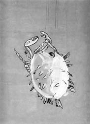
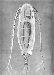
Figure: 4. Plankton. 7th grade, the topic "Crustaceans". Tinted paper (A3 format), chalk or white gouache, black ink, from satin
In the 8th grade, when studying fish, at the laboratory work "External structure of bone fish", we first draw an ordinary vobla, and then the children draw watercolors of representatives of different orders of fish from the magnificent color tables "Fishing fish" that we have at school.
Figure: 5. The skeleton of a frog. 8th grade, theme "Amphibians". Pencil, with a training drug
When studying amphibians, first - laboratory work "The structure of the skeleton of a frog", drawing in a simple pencil (Fig. 5). Then, after watching a short video clip, a watercolor drawing of various exotic frogs - leaf crawlers, etc. (We copy from calendars with high-quality photographs, fortunately, they are not uncommon now.)
With this scheme, rather boring pencil drawings of the same object are perceived as a normal preparatory stage for bright and individual work.
Important: technique
The choice of technique is essential to the successful completion of the job. In the classic version, you should take a simple pencil and white paper, but ... Our experience says that from the point of view of children, such a drawing will look unfinished, they will remain dissatisfied with the work.
Meanwhile, it is enough to make a pencil sketch in ink, and even take tinted paper (we often use colored paper for printers) - and the result will be perceived quite differently (Fig. 6, 7). The feeling of incompleteness is often created precisely by the lack of a well-developed background, and the easiest way to solve this problem is with tinted paper. In addition, using regular chalk or white pencil, you can almost instantly achieve the effect of flare or transparency, which is often needed.
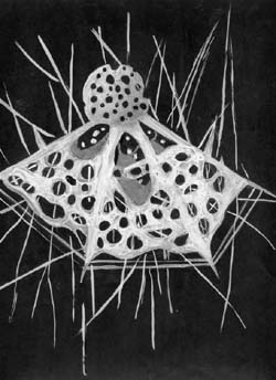
Figure: 6. Radiolaria. 7th grade, theme "The simplest". Tinted paper (A3 format) for watercolors (with a rough texture), ink, pastel or chalk, from satin

Figure: 7. Bee. 7th grade, topic "Insect orders". Ink, pen on pencil, volume - with a brush and diluted ink, fine details with a pen, from satin
If you find it difficult to organize work with mascara, use soft black liners or rollerballs (at worst, gel pens) - they give the same effect (Fig. 8, 9). Using this technique, be sure to show how much information is provided by using lines of different thickness and pressure - both to highlight the most important, and to create a volume effect (foreground and background). You can also use moderate to light shading.
Figure: 8. Oats. 6th grade, theme "Variety of flowering plants, Cereals family". Ink, tinted paper, from herbarium

Figure: 9. Horsetail and baloon. 6th grade, the topic "Spore plants". Ink, white paper, from herbarium
In addition, unlike classical scientific drawings, we often do work in color or use light toning to indicate volume (Fig. 10).

Figure: 10. Elbow joint. 9th grade, the topic "Musculoskeletal system". Pencil, with plaster cast
We tried many of the color techniques - watercolor, gouache, pastel, and eventually settled on soft colored pencils, only on rough paper. If you do decide to try this technique, there are a few important things to keep in mind.
1. Pick up soft quality pencils of a good company, for example, "Kohinoor", but do not give children a wide range of colors (basic enough): in this case, they usually try to pick up a ready-made color, which of course fails. Show how to achieve the correct shade by mixing 2-3 colors. To do this, you need to work with a palette - a piece of paper on which they select the desired combinations and pressure.
2. Rough paper will greatly facilitate the task of using light and strong colors.
3. Light short strokes should, as it were, sculpt the shape of the object: i.e. repeat the main lines (and not paint, contradicting the shape and contours).
4. Then the finishing touches are needed, juicy and strong, when the correct colors are already matched. It is often worth adding highlights, which will make the drawing very lively. The simplest thing is to use regular chalk (on tinted paper) or go through with a soft eraser (on white). By the way, if you use free-flowing techniques - chalk or pastel - you can then fix the work with hairspray.
When mastering this technique, you will be able to use it in nature, with a lack of time, literally "on your knee" (just do not forget about the tablets - a piece of packaging cardboard is enough!).
And, of course, for the success of our work, we definitely organize exhibitions - sometimes in the classroom, sometimes in the corridors of the school. Quite often, children's reports on the same topic, both oral and written, are timed to the exhibition. In general, such a project leaves you and the children with the feeling of a great and beautiful job that is worth preparing for. Probably, with contact and mutual interest with a drawing teacher, you can start work in biology lessons: analytical preparatory stage studying the object, creating a pencil sketch, and finishing it in the technique you have chosen together - in his lessons.
Here's an example. Botany, theme "Escape - bud, branching, structure of the shoot." A branch with buds is large in the foreground, in the background there are silhouettes of trees or bushes against a background of white snow and a black sky. Technique - black ink, white paper. Branches - from nature, silhouettes of trees - from photographs or book drawings. The title is "Trees in Winter" or "Winter Landscape".
Another example. When studying the topic "Insect orders", we do a short work "The shape and volume of beetles." Any technique that conveys light and shade and glare (watercolor, ink with water, brush), but monochrome, so as not to be distracted from the consideration and image of the form (Fig. 11). It is better to work out the details with a pen or a gel pen (if you use a magnifying glass, the legs and head will turn out better).
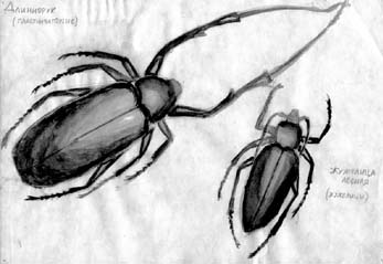
Figure: 11. Beetles. Ink, pen on pencil, volume - with a brush and diluted ink, fine details with a pen, from satin
Enough 1-2 beautiful works in a quarter - and drawing alive will delight all participants in this difficult process.