Telegram blocking and other symptoms of autoimmune Roscomencephalitis. Million Dollar Madness
I recently compared the actions of Roskomnadzor to block Telegram with an autoimmune disease: “Having recognized the imaginary terrorist threat, he went to fight with all the healthy tissues of the Internet.” Many wrote in the comments: “Lupus!” But no, this is not lupus. It is time to make a differential diagnosis and expand the biological metaphor of what is happening.
The lupus, so beloved by Dr. House's team, manifests itself in the form of a rash on the face, but in general it is a disease of connective tissue and its derivatives. If we imagine that people living in the country are the cells of a single organism, working collectively for its prosperity, then lupus is more likely when they infringe on the rights of truckers, fill people with garbage and do not repair roads.
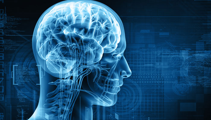
The Internet is a fiber of the country's nervous system. Therefore, massive blocking of sites should be considered autoimmune encephalitis. The disease is characterized by inflammation of the brain and the appearance of antibodies to molecules located on the surface of nerve cells. First of all, these are various neurotransmitter receptors. They are blocked by antibodies, like websites by IP addresses: in our country, the titer of such antibodies has already exceeded 18 million.
Since receptors of the same type are present in many nerve cells (only one cannot be blocked), the latter fail in groups. Please note that this is exactly what Roskomposor does!
Depending on which particular molecules become victims of a malfunctioning immune system, different variants of autoimmune encephalitis are observed. Each has its own set of symptoms, including psychiatric ones.
For example, we do not see the muscle stiffness syndrome characteristic of autoimmune encephalitis, with damage to GABA-A receptors or glycine, or movement disorders associated with damage to dopamine receptors. But one diagnosis fits perfectly.
The most common variant of autoimmune encephalitis is anti-NMDA receptor encephalitis (later we will return to what NMDA means). Psychiatric syndromes with NMDA receptor encephalitis include (I do not invent anything, everything is taken in the order indicated in the scientific publication):
1. Delusions of grandeur;
2. Paranoid delirium;
3. Hallucinations - sound and visual;
4. Unusual behavior;
5. Anxiety;
6. Fear;
7. Insomnia;
8. Confusion;
9. Loss of memory.
What, if not the paranoid delusions with hallucinations, is the round-the-clock search for fictional terrorists on Telegram? What, if not memory loss, blocking sites that store scientific and other valuable information, increasing censorship of the Internet? What, if not delirium of grandeur, ignoring the interests of millions of Telegram users and numerous organizations whose sites have laid down due to carpet locks? What, if not confusion, a denial of the absurdity of what is happening?
Coincidence? I don’t think so!

The symptoms of anti-NMDA receptor encephalitis are treated with immunosuppressants. That is, it is worth suppressing the activity of Roskomnadzor, recognizing that it is hyperactive. But, alas, everything is not so simple.
Everything is well provided for in the human body. Our brain is protected from the immune system - including the blood-brain barrier (in the metaphor with the Internet, anonymizers, Tor, VPN are trying to perform its function). Because for survival it is very important that nothing interferes with the transmission of information.
Therefore, autoimmune encephalitis is very rare. And, as a rule, they arise not just like that, but against the background of a tumor. In the case of anti-NMDA receptor encephalitis, it is usually ovarian teratoma (which makes sense, because Russia is female). Immature teratomas can sprout into nearby tissues and produce metastases.
The key characteristics of cancer cells are:
1. Violations of programmed cell death. In other words, the lack of cell turnover;
2. Violations of the control of the cell cycle;
3. Unjustified intake of glucose and other nutrients. Something like intercellular corruption. Together with offers to all other cells to tighten belts.
And again, all the signs are there. It is because of a corrupt tumor that the immune system does not solve real problems (for example, fire safety in shopping centers), but deals with inflammation of the brain. By the way, the NMDA receptor is a glutamate receptor. Therefore, it is not surprising that the deputies are very actively fighting this substance.
So, the final diagnosis: anti-NMDA receptor encephalitis in the presence of immature metastatic ovarian teratoma. Necessary tumor removal. Steroids to suppress the immune system. Intravenous immunoglobulin. Plasmophoresis to eliminate autoreactive antibodies. The main thing is that they do not have time to ban the import of drugs.
It remains to say that each of us can plant steroids
OBSERVATIONS FROM PRACTICE
UDC 615.06 + 616.831-002
MALIGNANT NEUROLEPTIC SYNDROME OR AUTOIMMUNE ANTI-SHYA RECEPTOR ENCEPHALitis? Fatal Case Study
DI. Malin, V.N. Gladyshev
Moscow Research Institute of Psychiatry - branch of FSBI
"The Federal Medical and Pharmaceutical Center named after V. P. Serbsky »Ministry of Health of Russia, Clinical Psychiatric Hospital No. 4 named after P. B. Gannushkina DZ Moscow
Malignant antipsychotic syndrome (ZNS) is a rare but extremely dangerous complication of antipsychotic therapy occurring with central hyperthermia, catatonic symptoms with muscle hypertonicity, impaired consciousness and a complex of somatovegetative disorders. The course of ZNS is accompanied by changes in the basic parameters of homeostasis and the function of vital organs and systems of the body and can lead to death. Mortality in ZNS according to various publications is from 5.5 to 10%, and the frequency of development is from 2 to 0.01% of all patients receiving antipsychotics. Most often, ZNS develops during neuroleptic therapy in patients with schizophrenia or schizoaffective disorder. In the world literature, cases of the development of complications in patients with affective disorders, dementia and organic psychoses are described. The development of ZNS can be noted when prescribing antipsychotics of various chemical groups, regardless of their dosages. Most often, the development of complications was noted with the appointment of a traditional antipsychotic - haloperidol. There are descriptions of the development of ZNS and the use of atypical antipsychotics - clozapine, risperidone, quetiapine and olanzapine, as well as against the background of the simultaneous cancellation of psychotropic drugs.
The etiology and pathogenesis of ZNS have not yet been fully studied. Most researchers attribute the development of the central nervous system to the blockade of dopamine receptors in the basal ganglia and hypothalamus, rather than the direct toxic effect of antipsychotics. In patients with ZNS, dopaminegric suppression and an increase in adrenergic and serotonergic activity are noted. A number of researchers consider ZNS as a manifestation of acute antipsychotic encephalopathy. In this case, signs of metabolism are detected on the EEG
encephalopathy with generalized inhibition of the electrical activity of the brain. The results of clinical and pathogenetic studies have established that immunological disorders and increased permeability of the blood-brain barrier, with neurosensitization of the body and subsequent autoimmune damage to the central nervous system, mainly the hypothalamus and visceral organs, play an important role in the pathogenesis of ZNS and febrile schizophrenia. Proof of this is the high humoral sensitization to various autoantigens of the brain with the detection of antibodies to the frontal lobe, optic tubercle and the maximum amount (up to 66%) to the hypothalamus. The cause of death is increasing disturbances of homeostasis and, first of all, water-electrolyte balance and hemodynamics, the phenomena of cerebral edema.
Analysis of pathomorphological changes in patients with fatal central nervous system in the world literature is not presented. The revealed pathological and morphological changes in the brain during febrile (hypertoxic) schizophrenia, and a number of researchers consider CNS as a form of lethal catatonia caused by antipsychotics (drug-induced), do not fit into any specific nosological form and can be attributed to the toxic-dystrophic process in combined with generalized discirculatory disorders. The following changes are detected in the thalamo-pituitary region of the brain in these patients: 1) acute swelling, vacuilization, ischemia and death of nerve cells; 2) swelling and swelling of the myelin sheaths of the gangliocytic fibers; 3) hypertrophy and degenerative changes of microgliocytes.
Risk factors for the development of ZNS are the presence of residual cerebral organic insufficiency in patients (transferred antenatal and perinatal hazards, craniocerebral trauma, infections and intoxications). Supposed
that physical exhaustion and dehydration arising against the background of psychomotor agitation can lead to increased sensitivity to antipsychotics and contribute to the development of central nervous system. The presence of catatonic disorders is also a risk factor for CNS.
Diagnosis of ZNS is based on the identification of the main symptoms of complications: central hyperthermia, catatonic symptoms with the development of stupor and muscle rigidity, impaired consciousness, as well as characteristic changes in laboratory parameters (moderate leukocytosis without a stab shift, leukopenia and accelerated ESR, sharp CPK activity in blood plasma).
The earliest sign of the development of ZNS in patients with schizophrenia and schizoaffective psychosis, important for the diagnosis of complications, is the appearance of extrapyramidal symptoms with an exacerbation of psychosis and the development of catatonic disorders in the form of a stupor with the phenomena of negativity and catalepsy. In this regard, some researchers consider ZNS as a neuroleptic variant of malignant or febrile catatonia, referring them to diseases of the same spectrum. This is confirmed both by the commonality of the clinical manifestations of febrile schizophrenia and central nervous system, and the similarity of biochemical and immunological disorders, as well as by the general principles of therapy. They include the abolition of antipsychotics, the appointment of tranquilizers, conducting infusion therapy and ECT. The effectiveness of the dopamine receptor agonist bromocriptine and dorethrolene myorelaxant in ZNS has not been confirmed by evidence-based studies. There is evidence of the effectiveness of plasmapheresis and hemosorption. The prognosis of the course of ZNS depends on how quickly neuroleptic therapy is canceled and intensive infusion therapy is prescribed that corrects homeostasis. With the timely abolition of antipsychotics, the adequacy of infusion therapy, and the differentiated use of ECT methods, it is possible to achieve a therapeutic effect in the majority of patients within the first 3-7 days, in accordance with the recommendations of B8M-5 ZNS, it is necessary to differentiate with such diseases as viral encephalitis, volumetric, vascular and autoimmune lesions of the central nervous system, as well as with conditions associated with the use of other drugs (amphetamines, phencyclidine, monoamine oxidase inhibitors, serotonergic other antidepressants and a number of other drugs).
In 2007, a series of cases were described for the first time, autoimmune KMBL receptor encephalitis occurring with psychotic symptoms and catatonia, autonomic disorders and hyperthermia, and the risk of death. Symptom
the math of this disease is similar to ZNS and febrile catatonia and causes difficulties in the differential diagnosis. The disease is caused by antibodies to QW1 and QW2 subunits of the glutamate KMBL receptor. Initially, anti-KMBL receptor encephalitis has been described in young women with ovarian teratomas. Subsequently, out of touch with the tumor process in people of both sexes and of different ages. Diagnosis of anti-MSCL receptor encephalitis is based on the detection of autoantibodies to QW1 and QW2 subunits of the glutamate KMBL receptor in plasma and cerebrospinal fluid. In recent years, cases of autoimmune encephalitis have been identified in patients with psychiatric hospitals with initial diagnoses of schizophrenia, schizo-affective disorder, narcolepsy, and major depressive disorder. Treatment of the disease involves immunotherapy with the appointment of immunoglobulin and methylprednisolone. The second-line drugs that are used in the absence of effect are rituximab in combination with cyclophosphamide. To stop psychomotor agitation, tranquilizers, atypical antipsychotics, or clompromazine can be used. There is positive experience with ECT and plasmapheresis.
Clinical case
Patient Sh., Born in 1988, was admitted for treatment to the Clinical Psychiatric Hospital No. 4 named after P.B.Gannushkina 06/18/2015, with a diagnosis of acute polymorphic psychotic disorder.
Anamnesis. The heredity of psychopathology is not burdened. Pregnancy and childbirth in the mother of the patient proceeded without pathology. Born on time. The eldest of 2 children. Has a younger sister. Previously, the development is correct. She was calm, balanced, sociable and active in character. She was ill with childhood infectious diseases without complications. I went to school from the age of 7. She studied well. She graduated from 9 classes of a secondary school, then a pedagogical college and a pedagogical institute. At the age of 22, she got married. She lived with her husband, a child of 3 years old from marriage, family relations are good. He works at the school as a primary school teacher. Has no bad habits. According to her husband, the patient’s mental state first changed since the beginning of June 2015. She became distracted, forgetful, anxious. Constantly asked her relatives: “did she feed the baby?”, “Did she go to the toilet”, said that she seemed to have “a head separate from the body”, sometimes unexpectedly fell to the floor, but immediately got up. 06.16.2015 applied for examination at the Federal State Budgetary Scientific Institution of the National Center for Neurology. Signs revealed on brain MRI
plot of gliosis in the right parietal lobe (8 mm-13 mm-18 mm), which must be differentiated from ischemic and demyelinating or volumetric process. Data for the presence of aneurysms, arteriovenous malformations at the studied levels have not been received. On the evening of the same day, she became anxious, restless, confused, asking "what is happening around?" There was a rise in systolic blood pressure to 180 mmHg. The night was restless. The next day, she began to express ridiculous ideas, believed that she was “bitten by a tick”, that she was pregnant. She claimed that songs sounded in her head. From time to time she felt fear, anxiety, was worried that she would not be able to work, thought that she would be “taken away from her child”, said “I will die”, noted that as if someone was controlling her, movements were happening against her will. 06/18/2015, she again turned to the center of neurology. She was excited at the reception, shouting “where is my mommy?”, Talking to herself, randomly waving her arms, growling, spitting. In connection with inadequate behavior, she was examined by a psychiatrist on duty and was hospitalized in PKB No. 4 in an involuntary manner.
Mental state at admission. Delivered to the department accompanied by orderlies with measures of physical restraint. Inspected within the bed. Productive contact is little available. Tense, anxious, listens to something, looks around. Responds only to whispering speech. Answers are quiet, concise, more often nods or shakes his head. From the conversation it is possible to reveal that she did not sleep for several nights, experiences the influx of thoughts in her head, the “sound of thoughts”. He does not deny the presence of “voices” that interfere with sleep and prohibit answering questions. Answers mainly "I do not know." At times she screams loudly, wriggles, spits.
Somatic condition: high growth, proper physique, satisfactory nutrition. The skin and visible mucous membranes of normal color. On the right elbow, there are traces of injections. On the face of a single red rash. Body temperature is normal. Zev is calm. In the lungs, vesicular breathing, no wheezing. NPV 16 min Heart sounds are muffled, rhythmic. Heart rate 82 beats / min. HELL 130/80 mm Hg The tongue is clean, moist. The abdomen on palpation is soft, painless. The liver and spleen are not palpable. The symptom of “generation” is negative on both sides. No swelling.
Neurological status: face symmetrical, pupils B \u003d 8, photoreaction saved. An increase in tendon reflexes is noted. Muscle tone is not increased. There are no meningeal signs, focal neurological symptoms are absent.
Laboratory examination data. The study of general clinical and biochemical blood and urine tests did not reveal any significant pathological changes, К ^ HIV, HBSAg, ISU -
negative, WB, BB - not identified. RPGA - tetanus - 0.77, diphtheria - 0.17. ECG - sinus rhythm, heart rate 55-62 per minute. Normal EOS.
Dynamics of the condition and ongoing therapy. From the first day of admission, the patient was prescribed haloperidol 15 mg / day i / m, trihexyphenidyl 6 mg / day, tiapride 400 mg / day i / m, chlorpromazine 25 mg / day i / m. Psychomotor agitation was stopped only in the evening. In the early days, the patient's condition remained unstable, there were episodes of psychomotor agitation with an influx of hallucinatory experiences, shouted incoherent phrases, talked to herself, lying in bed covered with a blanket with her head. I took treatment with duress, ate very little with persuasion. Productive contact remained unavailable. Gradually, psychomotor agitation was completely stopped during therapy. However, inhibition began to increase with an increase in muscle tone. All the time the patient lay motionless in bed, sometimes moving her lips. Answered only in a whisper. Symptoms of “wax flexibility” and “air cushion” appeared. In connection with the refusal of food, from June 23, 2015, infusion therapy with saline and glucose solutions up to 800 ml per day was prescribed. However, to improve the condition of the patient failed. On July 1, 2015, haloperidol and tiapride were canceled and olanzapine was prescribed at a dose of 20 mg / day, phenazepam 1 mg at night against the background of infusion therapy. After a relatively short period of improvement, when the patient began to independently move around the department and take food, deterioration occurred. From July 6, 2015, an increase in body temperature to 38.5 ° C and tachycardia to 110 beats began to be noted. in min, rigidity of the muscles of the lower and upper extremities, the phenomenon of catalepsy with the symptom of "air cushion" reappeared. High blood CPK values \u200b\u200b(2,427 units / L) were detected in the blood in a biochemical blood test, slight leukocytosis (8.4 thousand), and S0E 15 mm per hour. In order to exclude somatic pathology, the patient was repeatedly examined by a general practitioner: data for somatic pathology were not identified. On radiography of the lungs dated 07/14/2015, there were no pathological shadows. In order to prevent pneumonia, antibacterial therapy is prescribed - ceftriaxone at 1.0 v / m 2 times a day. On July 13, 2015, olanzapine was canceled and infusion therapy was increased to 1,200 ml / day. Despite the ongoing therapeutic measures, the condition remained serious. The patient lay in bed all the time, refused to eat, practically did not respond to treatment, sometimes responded only to whispering, symptoms of “wax flexibility” were noted, hyperthermia and muscle rigidity persisted. July 15, 2015 was examined by the emergency neurologist on duty.
Conclusion: the phenomena of cerebral edema on the background of intoxication syndrome. A CT scan of the brain, MRI with contrast, and transfer to a hospital with intensive care unit are recommended. At 19:50, accompanied by a resuscitation team, the patient was transferred to the PSO GKB them. S.P. Botkin to continue treatment and examination. Upon receipt, the condition was regarded as serious. Retardation with stunning elements persisted, did not respond to inverted speech, weakly responded to pain stimuli. An increase in tone in the muscles of the limbs and neck was noted. Inhibition, which was periodically replaced by excitement limited by the limits of the bed, with the repetition of individual words according to the type of speech stereotypes. In somatic status, pallor of the skin, tachycardia up to 110 beats were noted. in min., hyperthermia. For the differential diagnosis of demyelinating disease and encephalitis, lumbar puncture was performed - cytosis 40 in 3 ml, protein 0.33, lymphocytes 37, neutrophils 3. Antibodies to Epstein-barr virus, herpes virus, mycobacterium tuberculosis and to treponema pallidum were not found in cerebrospinal fluid. After examination by an infectious disease specialist, the diagnosis of viral encephalitis was withdrawn. On the MRI of the brain with contrast from July 21, 2015, an area of \u200b\u200bacute edema was detected in the semi-oval centers on the right. which should be differentiated with acute cerebrovascular accident according to the ischemic type, tumor, dimyelinating and autoimmune disease. The results of immunotyping of cerebrospinal fluid lymphocytes and lymphoproliferative disease have not been confirmed. In the intensive care unit, infusion therapy of up to 2 liters was carried out. per day under the control of diuresis, detoxification therapy, antibiotic therapy (cefritiaxon, amoxicycline). From July 24, 2015, dexamethasone 12 mg / day iv was added to the treatment regimen. Despite the therapy, the patient's condition remained severe, there was an increase in body temperature up to 40 ° C, a drop in blood pressure.
The conclusion of the consultation of doctors from 07.29.2015. The patient's condition is serious, febrile fever and catatonic symptoms persist. Most likely, the patient has febrile schizophrenia. The changes revealed on an MRI study, given their inconsistency of clinical symptoms, appear to be an accidental find and may be a consequence of a previous cerebrovascular accident.
07/29/2015, there was a respiratory arrest and cardiac activity. The started resuscitation measures did not lead to the restoration of breathing and cardiac activity. At 2215 hours biological death was observed.
At autopsy. Autoimmune encephalitis with a predominant lesion of the subcortical structures of the brain: the hippocampus, thalamus, hypothalamus. Perivascular lymphoplasmacytic infiltrates with the release of immunocompetent cells into the substance of the subcortical structures of the brain; perivascular and pericellular edema; dystrophy of gangliocytes with partial cytosis and reactive gliosis, with the formation of gliomesodermal foci. Cause of death: the patient's death (primary cause) occurred from autoimmune encephalitis, complicated by cerebral edema with dislocation of its trunk into a large occipital foramen (immediate cause of death).
Parsing. This clinical case demonstrates the difficulties of differential diagnosis and treatment of CNS. A 26-year-old patient developed an acute psychotic attack of a polymorphic psychopathological structure with acute sensory delusions, verbal pseudo-hallucinations and mental automatisms. From the first days of the manifestation, catatonic disorders in the form of impulsivity, negativism (responded only to whispering speech), and elements of hebephrenic excitation (growled, spat) were noted in the structure of the attack. Thus, the structure of psychosis was characteristic of manifest attacks traditionally described in schizophrenia and schizoaffective psychosis. Against the background of neuroleptic therapy with haloperidol and tiapride, there is an increase in inhibition with an increase in muscle tone, catalepsy with symptoms of “wax flexibility” and “air cushion” appears. The indicated transformation of psychosis is characteristic of the initial stage of development of the central nervous system. The abolition of haloperidol and tiapride and the appointment of the atypical antipsychotic olanzapine during infusion therapy only for a short time led to an improvement in the patient's condition. In the future, there is an increase in catatonic disorders - a stupor, alternating with excitation, somatic disorders appear in the form of hyperthermia, tachycardia, instability of blood pressure, characteristic changes in laboratory parameters (slight leukocytosis without a wand-nuclear shift, accelerated ESR and a sharp (10-fold) increase in CPK activity in blood serum). A thorough somatic, laboratory and instrumental examination, including a study of cerebrospinal fluid and MRI of the brain with contrasting, could not establish the cause that could underlie the development of a severe mental and somatic state. The patient died on the background of hyperthermia and the growing phenomena of cerebral edema, despite the abolition of antipsychotics, intensive care and the appointment of dexamethasone. The data of a pathoanatomical study revealed a manifestation of autoimmune encephalitis in a patient with a lesion
subcortical structures of the brain, which was the basis for the divergence of the diagnosis. At the same time, the patient was not tested for blood and cerebrospinal fluid to identify autoantibodies to KMBA receptors, on the basis of which autoimmune encephalitis is diagnosed. In addition, the results of a pathomorphological study do not contradict the diagnosis of ZNS, since the clinical and pathogenetic studies have proved the important role of autoimmune pathology with a primary lesion of the hypothalamus in the pathogenesis of the development of febrile seizures of schizophrenia. It is known that, when combined with blood plasma proteins, antipsychotics acquire the properties of haptens to which antibodies begin to form, blocking their antipsychotic effect. Under certain conditions, they are likely to provoke the development of an autoimmune process and cause the development of central nervous system. It should be noted,
that the algorithm for the diagnosis of ZNS until recently did not imply a study of blood and cerebrospinal fluid for the presence of autoantibodies to KMBA receptors. Moreover, in the world literature there are descriptions of cases when the originally diagnosed ZNS was revised after the detection of autoantibodies to KMBA receptors in the blood and cerebrospinal fluid. It can be assumed that early diagnosis of ZNS with the abolition of antipsychotics, the appointment of adequate infusion therapy and ECT would prevent a fatal outcome. However, the peculiarity of this case was that even before the manifestation of psychosis, the patient revealed changes in the MRI of the brain in the form of a portion of gliosis, which did not completely exclude the presence of a current organic central nervous system disease and diagnosed an endogenous disease - schizophrenia or schizoaffective psychosis based on the structure of psychopathological disorders.
LITERATURE
1. Avrutsky G.Ya. Paradise V. A. Tsygankov B. D. Clinic and course
malignant antipsychotic syndrome (acute febrile neuroleptic encephalopathy) // Zh. neuropathol. and a psychiatrist. 1987. Issue. 9.P. 1391-1396.
2. Govorin N.V., Lozhkina A.N. Antibodies to antipsychotics and their role
in the mechanisms of the formation of therapeutic resistance in psychopharmacotherapy of patients with paranoid schizophrenia // Zh. neuropathol. and psychiatry. 1991. Vol. 7, T. 91.S. 117-121.
3. Kekelidze Z.I. Chekhonin V.P. Critical conditions in psychiatry.
M .: GNTSSSP im. V.P. Serbsky, 1997.362 s.
4. Malin D.I. The effectiveness of plasmapheresis in the treatment of side effects and complications of antipsychotic therapy // Social and clinical psychiatry. 1993. No. 4. P. 82-84.
5. Malin D.I. Side effect of psychotropic drugs. M .:
University book, 2000.207 p.
6. Malin D.I., Ravilov R.S., Kozyrev V.N. The effectiveness of bromocriptine and dantrolene in the treatment of malignant antipsychotic syndrome // Russian Psychiatric Journal. 2008. N ° 5.P. 75-81.
7. Romasenko V.A. Hypertoxic schizophrenia. M .: Medicine,
8. Tiganov A.S. Febrile schizophrenia: clinic, pathogenesis, treatment.
M .: Medicine, 1982. 128 p.
9. Tsygankov B.D. Clinical and pathogenetic patterns of development
febrile seizures of schizophrenia and their treatment system. - M .; -1997.- 232c.
10. Chekhonin V.P., Morozov G.V., Morkovkin V.M., Kekelidze Z.I. Immunochemical study of the permeability of the blood-brain barrier in critical conditions due to febrile schizophrenia and acute alcoholic encephalopathy // Mat. 8th congress neuropath. and a psychiatrist. M., 1988. T 3.P. 132-134.
11. Agrawal S., Vincent A., Jacobson L. et al. Successful treatment of antiN-methyl-d-aspartate receptor limbic encephalitis in a 22-monthold child with plasmapheresis and pharmacological immunomodulation // Arch. Dis. Child 2009. Vol. 95. P. 312.
12. Braakman H.M., Moers-Hornikx V.M., Arts B.M. et al. Pearls and oysters: electroconvulsive therapy in anti-NMDA receptor encephalitis // Neurology. 2010. Vol. 75. P. 44-46.
13. Caroff S.N. The neuroleptic malignant syndrome // J. Clin. Psychiat. 1980. Vol. 41, N 3. P. 1-26.
14. Caroff S.N., Mann S.C. Neuroleptic malignant syndrome // Psycho-pharmacol. Bull. 1988. Vol. 24. P.25-29.
15. Consoli A., Ronen K., An-Gourfinkel I. et al. Malignant catatonia due to anti-NMDA-receptor encephalitis in a 17-year-old girl: case report // Child Adolesc. Psychiatry Ment. Health 2011. Vol. 5. P. 15.
16. Dalmau J., Tuzun E., Wu H.Y., Masjuan J. et al. Paraneoplastic anti-N-methyl-D-aspartate receptor encephalitis associated with ovarian teratoma // Ann. Neurol. 2007. Vol. 61. P. 25-36.
17. Dalmau J., Gleichman A. J., Hughes E.G. et al. Anti-NMDA receptor encephalitis: case series and analysis of the effects of antibodies // Lancet Neurology. 2008. Vol. 7, N. 12. R. 1091-1098.
18. Dalmau J., Lancaster E., Martinez-Hernandez, M. R. et al. Clinical experience and laboratory investigations in patients with anti-NMDAR encephalitis // Lancet Neurology. 2011. Vol. 10, N 1. P. 63-74.
19. Ghaziuddin N., Alkhouri I., Champine D. et.al. ECT treatment of malignant catatonia / NMS in an adolescent: a useful lesson in delayed diagnosis and treatment // J. ECT. 2002. Vol. 18, N 2. P. 95-98.
20. Gonzalez-Valcarcel J., Rosenfeld M.R., Dalmau J. Differential diagnosis of encephalitis due to anti-NMDA receptor antibodies // Neurología. 2010. Vol. 25. P. 409-413.
21. Keck P.E., Pope H.G., Cohen B.M., McElroy S.L., Nierenberg A.A. Risk factors for neuroleptic malignant syndrome. A case-control study // Arch. Gen. Psychiatry. 1989. Vol. 46. \u200b\u200bP. 914-918.
22. Keck P.E., Pope H.G., McElroy S.L. Declining Frequency of Neuroleptic Malignant Syndrome in a Hospital Population // Amer. J. Psychiatry. 1991. Vol. 148, N 7. P. 880-882.
23. Kiani R., Lawden, M., Eames P. et al. Anti-NMDA receptor encephalitis presenting with catatonia and neuroleptic malignant syndrome in patients with intellectual disability and autism // Br. J. Psych. Bull. 2015. Vol. 39. P. 32-35.
24. Kruse J.L., Jeffrey J.K., Davis M.C. et al. Anti-N-methyl-Daspartatere-ceptor encephalitis: a targeted review of clinical presentation, diagnosis, and approaches to psychopharmacologic management // Ann. Clin. Psychiatry. 2014. Vol. 26. P. 111-119.
25. Kuppuswamy P.S., Takala C.R., Sola C.L. Management of psychiatric symptoms in anti-NMDAR encephalitis: a case series, literature review and future directions // Gen. Hospital Psychiatry. 2014.P. 1-4.
26. Lancaster E., Dalmau J. Neuronal autoantigens - pathogenesis, associated disorders and antibody testing // Nature Reviews Neurology. 2012. Vol. 8, N 7. P. 380-390.
27. Lee A., Glick D., Dinwiddie S. Electroconvulsive therapy in a pediatric patient with malignant catatonia and paraneoplastic limbic encephalitis // J .ECT. 2006. Vol. 22. P. 267-270.
28. Lee E.M., Kang J.K., Oh J.S. et al. 18F-Fluorodeoxyglucose PositronEmission Tomography Findings with Anti-N-Methyl-D-Aspartate Receptor Encephalitis that Showed Variable Degrees of Catatonia: Three Cases Report // J. Epilepsy Res. 2014. Vol. 4, N. 2. P. 69-73.
29. Levenson J.L. Neuroleptic malignant syndrome // Am. J. Psychiatr5y. 1985. Vol. 142, N 10. P. 1137-1145.
30. Luchini F., Lattanzi L., Bartolommei N. et al. Catatonia and neuroleptic malignant syndrome: Two disorders of the same spectrum? Four case reports // J. Nerv. Ment. Dis. 2013. Vol. P. 36-42.
31. Mann S.C., Auriacombe M., Macfadden W. et all. Lethal catatonia: clinical aspects and therapeutic intervention. A review of the literature // Encephale. 2001. Vol. 27. P. 213-216.
32. Matsumoto T., Matsumoto K., Kobayashi T., Kato S. Electroconvulsive therapy can improve psychotic symptoms in anti-NMDA receptor encephalitis // Psychiatry Clin. Neurosci. 2012. Vol .66, N 3. P. 242-243.
33. Mirza M.R., Pogoriler J., Paral K. et al. Therapeutic Plasma Exchange For Anti-N-methyl-D-aspartate Receptor Antibody Encephalitis: A Case Report and Review of Literature // J. Clin. Apheresis. 2011. Vol. 26. P. 362-365.
34. Moscovich M., Novak F., Fernandes A. et al. Neuroleptic malignant syndrome // Arq. Neuropsiquiatr. 2011. N 5. P. 751-755.
35. Norgard N.B., Stark J.E. Olanzapine-associated neuroleptic malignant syndrome // Pharmacotherapy. 2006. Vol. 26. P. 1180-1182.
36. Patel A.L., Shaikh W.A., Khobragade A.K. Et al. Electroconvulsive Therapy in Drug Resistant Neuroleptic Malignant Syndrome // JAPI. 2008. Vol. 56. P. 49-50.
37. Reulbach U., Dutsch C., Biermann T. et al. Managing an effective treatment for neuroleptic malignant syndrome // Critical Care. 2007. Vol. 11. P. 4-10.
38. Sakkas P.I., Davis J.M., Janicak P.G., Wang Z.Y. Drug treatment of the neuroleptic malignant syndrome // Psychopharmacol. Bull. 1991. Vol. 27. P. 381-384.
39. Spivak B. Malin D., Kozirev V. et al. Frequencyensy of neuroleptic malignant
syndrome in a large psychiatric hospital in Moscow // Eur. Psychiatry. 2000. Vol. 15. P. 330-333.
40. Spivak B. Malin D., Vered Y. et al. Prospective evaluation of circulatory levels of catecholamines and serotonin in neuroleptic malignaut syndrome // Acta Psychitr. Scand. 2000. Vol. 101. P. 226-230.
41. Stauder K.N. Die todliche Katatonie // Arch. Psychiat. Nervenkr. 1934.
Bd. 102. S. 614-634.
42. Steiner J., Walter M., Glanz W. et al. Increased prevalence of diverse N-methyl-Daspartate glutamate receptor antibodies in patients with an initial diagnosis of schizophrenia: specific relevance of IgG NR1a antibodies for distinction from Nmethyl-D-aspartate glutamate receptor encephalitis // JAMA Psychiatry. 2013. Vol. 70. P. 271-278.
43 Strawn J.R., Keck P.E., Caroff S.N. Neuroleptic malignant syndrome // Am. . Psychiatry. 2007. Vol. 164. P. 870-876.
44. Titulaer M.J., McCracken L., Gabilondo I. et al., Treatment and prognostic factors for long-term outcome in patients with anti-NMDA receptor encephalitis: an observational cohort study // Lancet Neurology. 2013. Vol. 12, N 2. P. 157-165.
45. Trollor J.N., Sachdev P.S. Electroconvulsive treatment of neuroleptic malignant syndrome: a review and report of cases // Aust. NZ J. Psychiatry. 1999. Vol. 33. P. 650-659.
46. \u200b\u200bTsutsui K., Kanbayashi T., Tanaka K. et al. Anti-NMDA receptor antibody detected in encephalitis, schizophrenia, and narcolepsy with psychotic features // BMC Psychiatry. 2012. Vol. 12. P. 37.
MALIGNANT NEUROLEPTIC SYNDROME OR AUTOIMMUNE ANTI-NMDA
RECEPTOR Encephalitis? Fatal Case Study
DI. Malin, V.N. Gladyshev
The article is devoted to the analysis of a clinical case that ended in a fatal outcome, with a discrepancy between the clinical diagnosis and the data of the pathological study. It addresses the problem of the complexity of diagnosis and differential diagnosis of malignant antipsychotic syndrome and the so-called autoimmune
kMEL receptor encephalitis. A detailed consecration of the state of the problem under study with the analysis of modern publications is given.
Key words: lethal (febrile) catatonia, malignant antipsychotic syndrome, autoimmune UMBL receptor encephalitis, ECT, plasmapheresis, immunotherapy.
NEUROLEPTIC MALIGNANT SYNDROME OR AUTOIMMUNE ANTI-NMDA RECEPTOR ENCEPHALITIS?
Analysis of a fatal clinical case
D.I. Malin, V.N. Gladyshev
The authors analyze a fatal clinical case, when the pathologist "s investigation questioned the clinical diagnosis. The article discusses difficulties in diagnosis as well as differential diagnosis between neuroleptic malignant syndrome and the so called autoimmune NMDA receptor
encephalitis. The authors provide a state-of-the art description and a review of recent literature on the subject.
Key words: lethal (febrile) catatonia, neuroleptic malignant syndrome, autoimmune NMDA receptor encephalitis, ECT, plasmapheresis, immunotherapy
Malin Dmitry Ivanovich - Chief Researcher, Department of Therapy of Mental Illness, Moscow Research Institute of Psychiatry, a branch of the FSBI named after V.P. Serbsky "of the Ministry of Health of Russia; e-mail: [email protected].
Gladyshev Vitaliy Nikolaevich - Deputy Chief Physician of the Clinical Psychiatric Hospital No. 4 named after P.B.Gannushkina DZ of Moscow
In the medical literature, encephalitis refers to a whole group of diseases manifested by inflammatory processes of the brain. The disease has severe symptoms and can have a number of reasons, for example, an autoimmune process that causes anti-receptor encephalitis, or the presence of certain bacteria and viruses. Inflammatory processes of the brain require immediate qualified treatment, otherwise the risk of irreversible consequences or death is too high. This article will discuss anti-receptor encephalitis.
What is encephalitis?
Encephalitis causes various pathological disorders in the body and lead to the formation of dementia (dementia). The disease can affect not only the brain, but also part of the internal organs and joints.
Pathological conditions can be caused by a number of reasons. According to the factors provoking the disease, the following types of encephalitis are distinguished:
- inflammation caused by infection;
- bacterial or fungal encephalitis;
- a disease provoked by exposure to an organism of a toxic substance;
- autoimmune encephalitis.
The disease affects different parts of the brain. Inflammation can be localized in its cortex, subcortex, or cerebellum. Each type is distinguished by its own signs, symptoms and treatment methods. 
What is anti-receptor encephalitis? About it further.
Infectious and bacterial inflammation
Factors causing infectious encephalitis are viruses and bacteria. For example, herpes virus, HIV infection, encephalitis virus, tuberculosis bacteria, streptococcus and staphylococcus, toxoplasma. In addition, tick-borne encephalitis is a serious problem. This is a viral disease, the distributor of which are some types of ticks. The virus enters the body after an insect bite.
However, with tick-borne encephalitis, the brain is not always affected, in 50% of cases the patient experiences only fever. Japanese encephalitis also belongs to viral species. The disease is very dangerous and in most cases ends in death. This type of encephalitis is characterized by a rapid course, a few days after infection the patient falls into a coma. Herpes encephalitis is fatal in nine out of ten cases; it is practically untreatable.
How does anti-receptor encephalitis manifest? We will tell in more detail.
Autoimmune diseases

There is also a group of encephalitis that is caused by autoimmune processes in the body. In this case, the patient's own immune cells begin to attack the brain. Diseases of this nature are extremely difficult to treat, cause dementia, lead to impaired brain activity and the peripheral nervous system. In addition to dementia, the disease is accompanied by paralysis and seizures similar to epileptic. For diseases of this kind include, for example, limbic encephalitis. The disease causes an autoimmune response of the body to the presence of cancer cells or a disease of an infectious or viral nature. The rate of development of limbic encephalitis divides the disease into an acute and subacute form. The causes of anti-receptor encephalitis are discussed below.
Acute syndrome
In acute syndrome, the development of the disease occurs rapidly over three to five days. If you do not take urgent measures, then death occurs very quickly. With a subacute course of the disease, the first signs become noticeable after several weeks from the initial moment of development of the pathology. These conditions are characterized by the following symptoms:
- impaired memory;
- cognitive impairment;
- epileptic seizures;
- mental disorders (high level of anxiety, depression, agitation);
- behavioral disorders.
In addition, obvious signs are: progressive dementia, sleep disturbances, epileptic seizures with hallucinations. Cases when autoimmune brain lesions are correlated with the presence of cancer are very common. Typically, such encephalitis is caused by lung cancer. 
Anti NMDA Receptor Encephalitis
This is an autoimmune disease that affects young women more. In males, pathology is extremely rare. The features of this type of encephalitis include the presence of severe symptoms, which are expressed in serious psychoneurotic changes. That is why these patients are often diagnosed with schizophrenia instead of encephalitis. Women who were diagnosed with this pathology suffered from mental disorders (lack of coherent speech, impaired consciousness).
In addition, a characteristic symptom of anti-receptor encephalitis is a violation of short-term memory and muscle function. For example, in many patients there was an unreasonable contraction of the abdominal muscles, as well as jerky movements of the legs or arms.
About half of the examined patients had ovarian cancer. However, there may be cases when the patient does not have oncology. Moreover, there have been cases of the diagnosis of anti-receptor encephalitis in children not suffering from such diseases. Antibodies associated with certain brain structures called NMDA receptors spontaneously appear and begin to develop actively in them. Antibodies are fixed and block receptors, which in turn causes mental disorders, motor disorders and epileptic seizures. All this indicates that in many cases, doctors can not determine the exact cause of the disease. It should be noted that, in principle, they managed to identify this disease and learned to diagnose it no more than ten years ago. Symptoms and treatment of anti-receptor encephalitis are interrelated. 
Diagnostics
An experienced doctor who is not the first time encounters such pathologies will have suspicions even at the stage of examination of the patient. To make an accurate diagnosis, additional research is needed. As a rule, the appointment of magnetic resonance imaging is completely justified here. MRI will confirm or refute suspicions of inflammatory processes in the brain, however, it will not help to identify the cause of the disease.
For autoimmune diseases, including suspected anti-receptor encephalitis (we examined the causes of the disease), we analyze the presence of antibodies to the NMDA receptor. In some situations, cerebrospinal fluid analysis and a brain biopsy are prescribed. A biopsy is prescribed only as a last resort, when other methods to identify the cause of the disease are not informative. In this case, you can not do without consulting an oncologist.
Possible complications
Autoimmune diseases are difficult to diagnose, therefore, in the absence of proper experience with the doctor, the patient may be in a psychiatric clinic due to an incorrect diagnosis. The lack of necessary treatment leads to psychiatric abnormalities, which are often irreversible. In addition, there is a high probability that the patient may fall into a coma. If the patient does not take the medications necessary for treatment, the vegetative state develops very quickly, and a third of the patients die. 
Treatment for anti-receptor encephalitis
To make a correct diagnosis, first of all, the patient is sent for examination and consultation to a neurologist. The disease is diagnosed in the presence of certain antibodies in the blood. To exclude an incorrect diagnosis, an oncologist examination is also required. With timely treatment and a well-structured oncological treatment, in most cases it is possible to achieve persistent and prolonged remission. Good results are also achieved with immunomodulators. But this type of treatment is available only if the suspicion of oncology turned out to be baseless.
To reduce psychiatric symptoms, patients are prescribed drugs with a sedative effect. They calm and normalize sleep. With the appearance and repeated recurrence of seizures, antispasmodic drugs are prescribed. Removal of acute inflammation is achieved using corticosteroids. They are administered intramuscularly, and the duration of the course of treatment is prescribed by the doctor.
Anti-prescription encephalitis is practically not completely cured. Treatment helps stop the further progression of the disease and eliminates the development of neurological disorders. If the disease was caused by oncology, then the elimination of the tumor gives a very stable result, and 70% of patients recover completely. How can brain anti-receptor encephalitis be prevented?

Prevention
Since childhood, we know that you need to go to the forest in closed clothing, which excludes ticks from exposed skin. Such measures help in the prevention of viral and bacterial encephalitis. It is also important to contact medical institutions in a timely manner and follow the instructions of doctors. As for brain diseases of an autoimmune nature, including anti-prescription encephalitis, the development of such pathologies cannot be prevented.
Conclusion
According to available data, almost half of patients suffering from anti-receptor encephalitis fully recover. A third of patients have mild residual effects, and a small proportion of patients suffer from serious complications. About 10% of patients died.
Therefore, we must once again emphasize that when a tumor is detected at an early stage and its removal, the body functions are restored in full, that is, recovery occurs. All this allows us to conclude that it is necessary to consult a doctor at the first symptoms of the disease in order to increase the chance of a successful outcome.
The famous German philosopher Arthur Schopenhauer claimed that nine-tenths of our happiness depends on health. Without health - there is no happiness! Only complete physical and mental well-being determine human health, help us successfully cope with illnesses, adversities, lead an active social life, reproduce offspring, and achieve our goals. Human health is the key to a happy, fulfilling life. Only a healthy person in all respects can be truly happy and able toto the fullest extent, feel the fullness and diversity of life, experience the joy of communicating with the world.
They say that cholesterol is so unflattering that it’s just right for them to scare children. You should not think that it is a poison which only does that destroys an organism. Of course, it can be harmful, and even be dangerous to health. However, in some cases, cholesterol is extremely necessary for our body.
The legendary balm “asterisk” appeared in Soviet pharmacies in the 70s of the last century. It was in many ways an indispensable, effective and affordable drug. They tried to treat everything in the world with an “asterisk”: ARI, insect bites, and pain of various origins.
Language is an important organ of a person that can not only talk incessantly, but without saying anything, can tell a lot. And there’s something to tell him, especially about health.Despite its small size, the language performs a number of vital functions.
Over the past few decades, the prevalence of allergic diseases (AZ) has gained epidemic status. According to recent data, more than 600 million people worldwide suffer from allergic rhinitis (AR), approximately 25% of them in Europe.
For many people, there is an equal sign between a bathhouse and a sauna. And a very small number of those who realize that the difference exists can easily explain what the difference is. Having examined this question in more detail, we can say that there is a significant difference between these pairs.
Late autumn, early spring, periods of thaw in the winter season - this is a period of frequent colds, both adults and children. From year to year, the situation repeats itself: one member of the family gets sick and behind it, as in a chain, everyone suffers a respiratory viral infection.
Some popular medical weeklies can read odes to salou. It turns out that it has the same properties as olive oil, and therefore it can be consumed without any reservations. At the same time, many argue that you can help the body "cleanse" only by starvation.
In the 21st century, thanks to vaccination, it has decreased significantly prevalence infectious diseases. According to WHO, vaccination prevents two to three million deaths per year! But, despite the obvious benefits, immunization is shrouded in many myths that are actively discussed in the media, and indeed in society.